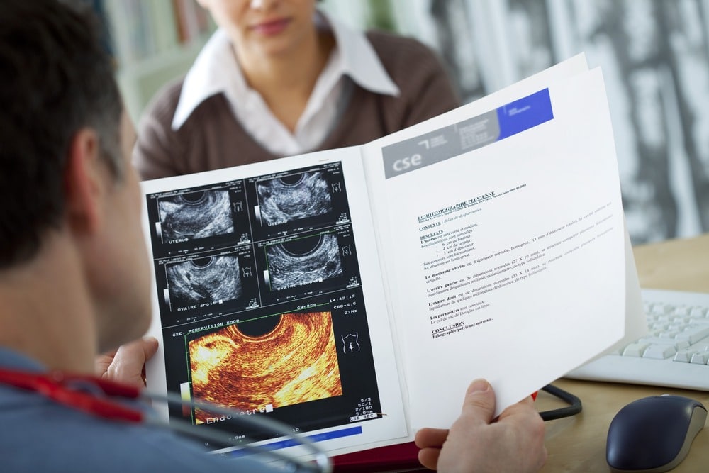
Ultrasound plays a vital role in reproductive health, especially in assessing and tracking the menstrual cycle for women aiming to understand their fertility better.
This method, often referred to as an “assessment” or “tracking cycle,” involves a series of ultrasounds timed within the menstrual cycle to observe follicle development, ovulation timing, and endometrial changes.
Such tracking can reveal key insights about a woman’s reproductive health, including issues like irregular ovulation, hormonal imbalances, or conditions like polycystic ovary syndrome (PCOS).
By following the cycle in real-time, both patients and healthcare providers gain critical information that can support natural conception or guide assisted reproductive treatments.

The Purpose of an Assessment or Tracking Cycle
Assessment and tracking cycles serve as a detailed, individualized method for observing a woman’s ovulatory pattern over a single or multiple menstrual cycles. For those seeking to conceive, the goal of this monitoring is to pinpoint the most fertile days with accuracy, enhancing the likelihood of conception. Beyond conception, tracking cycles reveal vital information about the ovulation process and overall cycle health. This detailed ultrasound monitoring proves valuable for a range of patients, from those with irregular cycles to individuals experiencing unexplained infertility or age-related changes in fertility.
Specialists in fertility and reproductive endocrinology rely on this method as an objective and precise tool to document and evaluate each stage of the cycle, from follicular growth to endometrial readiness. According to research in the Journal of Obstetrics and Gynaecology, monitoring cycles with ultrasound offers “an unparalleled view into ovarian activity and follicular dynamics” which can directly inform both natural and assisted conception efforts.
Components of the Tracking Cycle: Understanding What We Monitor
Follicle Development: Tracking the Foundation of Fertility
The growth and maturation of ovarian follicles is essential to successful ovulation. Follicles are fluid-filled sacs in the ovaries, each containing an immature egg. During a menstrual cycle, several follicles may begin to grow under the influence of follicle-stimulating hormone (FSH), but typically, only one reaches full maturity. This dominant follicle then releases an egg during ovulation.
Ultrasound allows us to monitor the growth of these follicles, measuring their size and progress at specific points within the cycle. Typically, ultrasound scans are performed around day 10 of the cycle and then every 2-3 days until ovulation. This sequence of scans provides detailed data about the rate of follicular growth, the size of the dominant follicle, and, ultimately, the timing of ovulation.
In clinical practice, follicles ideally reach about 18-25 mm in diameter before ovulation. According to a study published in Fertility and Sterility, the tracking of follicular development through ultrasound has a high predictive value for pinpointing ovulation within a 24-hour window. This level of accuracy is particularly beneficial for timing intercourse or medical interventions like intrauterine insemination (IUI).
Endometrial Thickness and Pattern: Preparing for Implantation
Alongside follicular tracking, ultrasound assessment of the endometrium (the uterine lining) is a key component of the tracking cycle. The endometrium undergoes a series of changes throughout the menstrual cycle, thickening in response to estrogen and maturing under the influence of progesterone after ovulation. A well-developed endometrial lining is essential for successful implantation of a fertilized egg.
Studies published in Human Reproduction Update highlight the importance of endometrial thickness for implantation success, noting that an optimal endometrial thickness ranges from 7 to 14 mm around the time of ovulation. Monitoring this thickness, as well as the endometrial pattern (triple-line or homogeneous), helps predict the uterus's receptivity to an embryo. For patients undergoing fertility treatments, such as IVF, endometrial assessment informs decisions about the timing of embryo transfer, enhancing implantation rates.
Hormonal Influence and Ovarian Reserve Evaluation
The interplay between hormones like FSH, luteinizing hormone (LH), estrogen, and progesterone drives the menstrual cycle. Ultrasound tracking, often paired with blood tests, provides a dynamic view of how these hormones impact follicular development and endometrial changes in real time. This combination reveals information about ovarian reserve, particularly in women over 35 or those with low ovarian reserve concerns.
Ovarian reserve refers to the quantity and quality of a woman’s remaining egg supply. By tracking how ovaries respond to FSH, and measuring antral follicles (small follicles present at the start of the cycle), ultrasound offers a glimpse into ovarian reserve. According to the Journal of Clinical Endocrinology & Metabolism, low ovarian response may indicate diminished ovarian reserve, which can affect both natural fertility and the success of assisted reproductive technologies (ART).

The Ultrasound Process in Tracking Cycles: What to Expect as a Patient
Scheduling and Timing of Ultrasound Appointments
Tracking cycles require precision in scheduling. The typical monitoring sequence begins with a baseline scan around day 3 of the menstrual cycle to assess baseline follicular activity and measure antral follicles. The second scan occurs around day 10, followed by scans every 2-3 days until ovulation. In certain cases, additional post-ovulation scans may be conducted to evaluate the corpus luteum and confirm ovulation.
A common technique used for these evaluations is the transvaginal ultrasound, which offers a closer and clearer view of the ovaries and uterus compared to abdominal ultrasound. This method enhances detail, allowing radiologists to accurately assess follicle size, endometrial pattern, and other structural factors.
What Radiologists Look For in Each Scan
Each ultrasound session in the cycle tracking process provides specific insights. During the initial scan, the radiologist examines the ovaries for the number and size of antral follicles, which can indicate ovarian reserve. The follow-up scans focus on the growth of the dominant follicle, while simultaneously tracking endometrial changes. Radiologists also observe blood flow patterns to follicles, which provide additional clues about follicular health and the likelihood of ovulation.
These details support fertility treatment plans and inform patients about their optimal conception days. By observing the gradual progression, radiologists can accurately predict ovulation, a critical factor in timing interventions or planning natural conception.
Interpreting Results: Common Findings and Their Implications
Normal Follicular Development
In a typical cycle, a dominant follicle grows from an initial size of about 4-6 mm to 18-25 mm by the time of ovulation. This progression is a strong indicator of a healthy ovulatory cycle. If a dominant follicle reaches maturity and ruptures, it releases an egg, confirming ovulation has occurred. Observing this pattern consistently over a few cycles can reassure patients about their reproductive health.
Abnormal Follicular Development
Irregularities in follicular development can suggest hormonal imbalances or ovarian conditions like PCOS. In patients with PCOS, multiple small follicles may form, but they often fail to mature and release an egg, leading to anovulatory cycles. Research from The Lancet reports that tracking cycles in women with PCOS often reveals “arrested” follicles that do not progress past a certain stage, helping diagnose the condition.
Endometrial Abnormalities and Their Impact on Fertility
Endometrial issues, such as a thin or irregularly developed lining, can hinder implantation even if fertilization occurs. A thin lining may indicate inadequate estrogen stimulation, while a delayed development pattern may suggest a hormonal imbalance in the luteal phase. Conditions like luteal phase insufficiency, characterized by low progesterone levels, prevent proper endometrial maturation, reducing implantation chances.
Studies in the Journal of Assisted Reproduction and Genetics indicate that tracking cycles provide early detection of luteal phase issues, allowing for hormonal treatment interventions that can improve implantation rates.
Supporting Fertility Treatments with Tracking Cycles
Cycle tracking is a cornerstone of many fertility treatment plans, especially those involving ART like IUI and IVF. With precise timing, ultrasound tracking can identify the ideal window for interventions, optimizing success rates. By aligning insemination or embryo transfer with peak endometrial receptivity and follicular readiness, tracking cycles improve treatment efficacy and outcomes.
Even for couples pursuing natural conception, monitoring through ultrasound supports more accurate timing of intercourse. A publication in Reproductive Biomedicine Online highlights that patients who use tracking cycles to time natural conception have significantly higher pregnancy rates than those who rely solely on home ovulation tests.
Frequently Asked Questions
Why are multiple scans needed within one cycle?
Multiple scans capture the dynamic changes of the menstrual cycle, ensuring accurate tracking of follicular growth and endometrial development. This step-by-step monitoring is essential for identifying the peak fertility window, especially for patients with irregular cycles.
How accurate is ultrasound in predicting ovulation?
Ultrasound tracking offers high accuracy, with studies showing it can predict ovulation within a 24-hour window, making it one of the most reliable methods for timing fertility-related interventions.
Can a tracking cycle diagnose underlying fertility issues?
Yes, tracking cycles reveals valuable insights about ovulation patterns, follicular health, and endometrial receptivity. These insights can help diagnose hormonal imbalances, anovulation, or luteal phase defects that may impact fertility.
What does it mean if follicles do not develop as expected?
Poor follicular development can indicate hormonal imbalances or ovarian issues, which can affect ovulation and fertility. Early detection through tracking allows for timely intervention with potential treatments.


