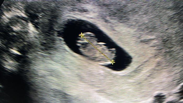
Your eight-week private ultrasound scan can be an exciting and apprehensive moment.
If this is your first pregnancy ultrasound you will be understandably anxious as getting a glimpse of your baby for the first time is a big deal.
Why have an 8-week baby ultrasound scan?
From anywhere between 8 and 12 weeks pregnant, your healthcare professional might suggest that you schedule your first ultrasound appointment. This is also called your “dating” scan.
The main reason for this scan is to confirm the gestational age of your baby. This date is based on your baby’s size (Crown Rump Length or CRL) and will be a very close estimation.
Some eight-week ultrasounds might be performed for other reasons, including:
- If you are experiencing bleeding
- If multiple pregnancies are suspected
- To check the size of your embryo
- To confirm that your baby has a heartbeat
- To check the health of your ovaries and fallopian tubes
- To rule out an ectopic pregnancy or other problems
If you have just discovered you are pregnant, but you aren’t sure when you became pregnant your chosen healthcare professional might also recommend an early pregnancy ultrasound scan.
Eight weeks pregnant is an early stage to perform an ultrasound, and you wouldn’t ordinarily need one so early.
What to expect during your 8-week ultrasound
Depending on your healthcare professional and your personal preferences, your eight week ultrasound can be performed using an ultrasound probe across your abdomen or another type of probe inside your vagina. A vaginal ultrasound is helpful if your bladder isn’t full enough or your uterus is still too small to see.
At this point in your pregnancy, an ultrasound can confirm that your baby is healthy and progressing as it should be.
If you are having twins (or more), you might be able to see multiple yolk sacs and multiple heartbeats. However, as it is still early days one baby may be missed at this stage.
Don’t be afraid to ask your sonographer questions. If this is your first ultrasound it’s very normal that you would be curious about what you see on screen.
Your baby at 8 weeks
At eight weeks pregnant, your baby will measure about 1.6 centimetres and it will be losing its little tadpole tail. Keep in mind that your baby’s big forehead and tiny body will still make him or her look rather disproportionate at this stage.
Your little one will also start to make involuntary movements, similar to a slight flicker or a jump. These movements are extremely tiny so you won’t be able to feel them.
Inside and out, a number of your baby’s body parts will start to become more defined as well, including their:
- Nose
- Lips
- Eyelids
- Arms
- Legs
- Bones
- The valves and air passages in their heart
Every pregnant woman is offered ultrasound scans during pregnancy. However, the number and timeline will be different for each woman.

Your ultrasound schedule will depend on a few key factors, including:
- The progression and health of your pregnancy
- Your personal preferences
- Your chosen healthcare professional
- Whether you will be giving birth in a public or private hospital
- Your medical history
If you didn't have an ultrasound last week, your nine-week ultrasound will likely be scheduled to assess the gestational age of your baby. If you're not sure when your last menstrual period (LMP) was, a scan at nine weeks will be able to confirm your approximate date of conception.
Your first ultrasound can be a very emotional experience so it's a good idea to take along your partner or close family member for support.
The purpose of a 9-week ultrasound
Depending on your unique pregnancy, your chosen healthcare professional may schedule a private baby ultrasound scan at nine weeks for a few different reasons.
If this is your first ultrasound, it will give you the opportunity to accurately determine your due date. Especially if you haven't tracked when your LMP was.
Knowing how far along you are in your pregnancy is important. At some point between 11 and 13 weeks, your healthcare professional will suggest conducting a Nuchal Translucency (NT) scan. This scan tests for Down syndrome and for accurate results, you need to know how far along you are.
If you have miscarried a previous pregnancy or you have experienced some level of vaginal bleeding over the last nine weeks, you may also be offered an ultrasound. This scan can confirm whether your pregnancy is progressing healthily.
What to expect during your 9-week ultrasound
This ultrasound may be conducted vaginally or externally on your abdomen. Know that if your healthcare professional has officially referred you for an early scan your insurance will probably cover it but please first check with your provider.
At nine weeks, you will be able to see your baby's head, body and limbs. You will also be able to hear your little one's heartbeat for the first time with a Doppler monitor. Bring some tissues with you; this can be a very emotional moment.
It's also important to understand that miscarrying during the first three months of pregnancy is quite common.
If your ultrasound shows that your baby is growing slowly, or has a lower than average heartbeat your chances of miscarrying are high. If you have been experiencing pain or vaginal bleeding, you might be somewhat prepared for this news. However, no matter how prepared you think you may be, hearing your miscarriage suspicions confirmed is likely to be a distressing experience.
Your baby at 9 weeks
At nine weeks, your baby will measure approximately 2.5 centimetres. The foetus will resemble a green olive and weigh less than 2 grams.
Your little one's eyes will have grown larger and even have some colour, but their eyelids will still be fused shut. Your ultrasound may be able to show you the beginnings of what will be your little one's fingers and toes too.
If you have not yet had any type of pregnancy ultrasound and you are around 12 weeks pregnant, your maternity care provider may suggest you have one. There are many reasons for having an ultrasound at this stage, but one of the most common is to screen for one of the congenital chromosomal abnormalities – Trisomy 21, otherwise known as Down Syndrome. This means that there is an extra chromosome – 21 contained in every cell of the body.
People with Down Syndrome have physical and intellectual disabilities. Older women are more at risk of having a baby with Down Syndrome.
When a woman is 12 weeks pregnant her risk of having a baby with Down Syndrome can be fairly accurately assessed. When a foetus has Down syndrome they tend to have more fluid at the base of their neck, in the region known as the nuchal fold area. This fluid can be measured in a test called nuchal translucency. A foetus with Down Syndrome has a measurement which is thicker than in those who do not.
It is worth remembering though, that a larger than average nuchal fold measurement is not a guarantee that the baby will have chromosomal problems. Other tests for Down Syndrome need to be done if in doubt. In addition, blood tests such as a Chorion Villus Sampling test or an amniocentesis help to clarify any suspicions.
I can see my baby!
The 12 week ultrasound may be the first time parents have seen their little baby. So this is exciting if a little nerve-wracking time. It's completely normal for parents to consider the possibility that their baby may not be developing as it needs to and perhaps build apprehension before the procedure. After all, this is one of the reasons why a 12-week ultrasound is recommended.
One of the benefits of having an ultrasound so early in pregnancy is that if complications are found, then parents may be given a choice of continuing with the pregnancy or not. Medical recommendations on this issue are very important. Ethical, religious and personal belief systems also need to be carefully balanced and weighed up.
Parents need to feel as if they are fully informed and comfortable with the explanations provided by the sonographer doing the 12-week ultrasound. Follow up care by the healthcare team are equally as important.
What is a first-trimester screening test?
Maternity care providers will often suggest a pregnant mother has a blood test taken when she is 10 weeks pregnant. This is specifically to conduct a measurement of pregnancy hormones. If chromosomal abnormalities are present, these results can be out of the normal range.
These blood tests, in combination with the 12-week ultrasound, provide what is known as “A First Trimester Screening Test”. A mother's age, including the results from the blood test, and the findings of the ultrasound all provide an individualised picture of her risk of having a baby with Down Syndrome. This screening test is not a definite diagnosis of chromosomal problems, but rather provides a risk assessment. If there are concerns, then further testing can be done.
How will they do the 12-week ultrasound?
The 12-week ultrasound is generally done via the mother's abdomen. It's not always necessary to have a full bladder, however, the individual sonographer may recommend that you have a partially full bladder. This will help to lift your uterus up out of your pelvis so it is easier to see the foetus. Sometimes it is necessary to do a vaginal ultrasound. This will lead to even clearer images.
Reasons to have a 12-week ultrasound
- To check that the foetus is developing as it should be. The measurements of the foetus's skull – the Biparietal distance is calculated and compared against standard lengths for foetuses at similar gestational ages.
- To see if the foetus has a heartbeat. This should be clearly detectable at the 12-week ultrasound.
- To confirm pregnancy dates and estimate the date of delivery.
- To check for multiple foetuses and confirm if one or more is present.
- To check the size of the foetus and developing placenta.
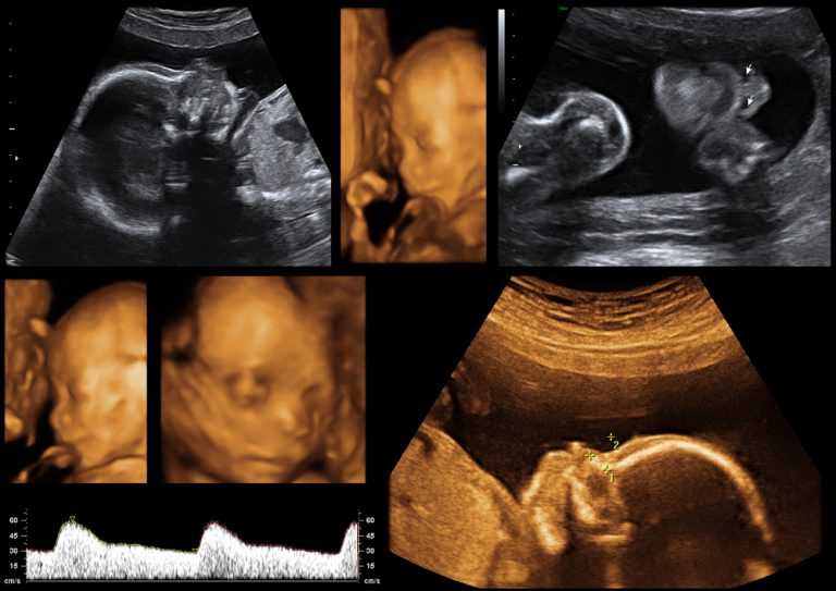
- To measure the amount of fluid at the base of the foetus's neck and make an individualised risk assessment of them having Down Syndrome. The sound waves from the ultrasound return echo-free measurements. This is because of space which is translucent due to its fluid content.
- To check for other physical abnormalities in the foetus.
- To check the uterus, fallopian tubes and pelvic region for other complications.
What else is measured during a 12-week ultrasound?
- The foetus's length, specifically from its head to its bottom. This is known as a Crown Rump length.
- A general check of the mother and foetus's internal organs and structures.
Many parents are amazed by the amount of detail they can see at the 12-week ultrasound. They are also surprised by their foetus's movements and agility. Of course, at 12 weeks gestation, it is too early for a pregnant mother to be aware of her baby moving. And it can be a strange sensation when looking at the monitor and seeing movement but not being able to physically detect it.
Many parents feel an instant emotional connection with their baby when they see it for the first time. It's not uncommon for fathers to say that the whole pregnancy idea was a little foreign and somehow not real. But being able to see their baby rather than talking about it and having to use their imagination, makes all the difference.
When will I know if everything's alright with my 12-week ultrasound?
You should be told straightaway if everything is going well. If you have had your biochemistry blood tests taken before the ultrasound and these results are back, then you should be able to have these results as well straight after your ultrasound is finished. Many maternity healthcare providers recommend mothers have the blood test at 10 weeks gestation.
The sonographer will be able to talk their way through the procedure and turn the screen so that you can see what they are looking at during the ultrasound. There may also be a separate monitor for you and your partner to look at. If you want an explanation, then just ask the sonographer to tell you what they are checking.
If they are unsure or want clarification they will often request a colleague come into the room and have a look at the ultrasound. Obviously, this can be a pretty unnerving process especially if you've not had any reason to believe that there are any complications.
How accurate is the first-trimester screening test?
At the current time, the combined First Trimester Screening is thought to be the most accurate test for Down Syndrome. For those women whose results return a high risk of carrying an embryo with Down Syndrome, the next stage is generally Chorionic Villus Sampling or an Amniocentesis.
Having a low-risk result for the First Trimester Screening Test does not give a 100% guarantee that there will not be a chromosomal abnormality. What it does is categorise a pregnancy into an increased or decreased risk.
How long will my 12-week ultrasound take?
Generally, bookings of 30 minutes are made. This allows the sonographer enough time to do a thorough and comprehensive check and assessment. Try not to squeeze your appointment time between a lot of other tasks you need to achieve in the same day.
Put aside some time before and after your ultrasound so you can make it to the appointment in plenty of time, and have the chance to reflect on it afterwards. Ask your partner to be with you on the day, and aim to enjoy this as an event you can both share.
Some couples choose to bring their parents along as well and view this as an opportunity to meet their grandchild for the first time. How you manage this is your choice, just be mindful that ultrasound rooms can be quite small, so accommodating more than a couple of people can present a practical challenge.
Are 12-week ultrasounds part of my routine pregnancy care?
Ultrasounds during pregnancy are routinely offered because they provide such an excellent means of diagnosing problems if they are present. They are also low risk, non –invasive and relatively low cost considering the amount of information they give. But you are entirely free to make your own choices regarding whether you want to have pregnancy ultrasounds or not.
Some parents feel very strongly that having an ultrasound is not right for them. Part of their reasoning is that if abnormalities are found, then they may be put into a position of having to make decisions based around these findings.
Some parents choose to wait until the screening ultrasound at 18-20 weeks and feel that at 12 weeks it is still too early to be able to see much of their baby's development. If you are in any doubt, then speak with your maternity care provider about your own individual needs.
Medically Reviewed by Tareq Ismail Pg(Dip), BSc (Hons)
[post-views]
Medically Reviewed by Tareq Ismail Pg(Dip), BSc (Hons)


