Introduction to Ultrasound Scanning
Ultrasound scanning, a cornerstone of modern medical diagnostics, is a fascinating blend of science and technology that has revolutionized the way we view the human body. This technique uses high-frequency sound waves to create live images from inside the body, providing a window into our inner workings without any invasive procedures.
The beauty of ultrasound lies in its simplicity and safety. Unlike other imaging techniques, it doesn’t rely on ionizing radiation, making it a preferred choice for a variety of medical applications. Its non-invasive nature is particularly crucial in obstetrics, where it plays a vital role in monitoring the health and development of the fetus. As described in a publication in the Journal of Ultrasound in Medicine, ultrasound has been instrumental in evaluating pathologic pulmonary conditions, showcasing its versatility beyond just prenatal care.
Ultrasound’s application extends well beyond obstetrics, touching various medical fields. For instance, it’s routinely used in cardiology to assess heart function, as highlighted in research published in Interface Focus. In the realm of sports medicine, studies from the American College of Sports Medicine have underscored its value in diagnosing muscle and tendon injuries. Moreover, a study conducted at Peking University entitled “Effects of Dehydration and Rehydration” utilized ultrasound technology to gain insights into bodily changes under extreme conditions.
The technology behind ultrasound is continually advancing, with newer studies like those published in the European Journal of Nutrition exploring its evolving applications. These advancements not only enhance diagnostic accuracy but also open doors to new possibilities in medical imaging.
Ultrasound scanning represents a remarkable intersection of technology and healthcare. Its ability to provide real-time, detailed images of the body’s internal structures, as outlined in a study published in BMA by N. Smith et al in 2019, has made it an indispensable tool in modern medicine. As we continue to delve into the capabilities of this technology, ultrasound stands as a testament to how far medical imaging has come, offering both patients and physicians a safe, reliable, and non-invasive diagnostic option.
Principles of Ultrasound Imaging
Ultrasound imaging, also known as ultrasound scanning or sonography, is a widely used diagnostic tool in the medical field. This technology employs high-frequency sound waves to create images of structures within the body. It is favored for its non-invasive nature, lack of ionizing radiation, and real-time imaging capability. Let’s delve into the fundamental principles that govern ultrasound imaging.
How Ultrasound Works
Ultrasound imaging starts with the transmission of high-frequency sound waves into the body using a small probe called a transducer. As these sound waves travel through the body, they encounter various internal structures, which reflect the waves back to the transducer. The returned echoes are then captured and transformed into visual images. This process is remarkably safe, as highlighted in a study published in the journal “Interface Focus” by G. ter Haar, which emphasizes the safety of diagnostic ultrasound in various clinical applications, including monitoring pregnancy and investigating heart function.
Types of Ultrasound Imaging
There are several modes of ultrasound imaging, each serving a specific diagnostic purpose. The most common types include:
- B-mode Ultrasound: Provides a two-dimensional image of the internal structure of the body, useful for examining the fetus during pregnancy or organs like the liver and kidneys.
- Doppler Ultrasound: Measures and visualizes blood flow through arteries and veins, aiding in the detection of blood flow issues.
- 3D and 4D Ultrasound: Offers a more detailed and dynamic image, commonly used in obstetric ultrasounds to view the fetus.
Applications of Ultrasound
Ultrasound’s versatility allows for its application across various medical fields. A study from the “Journal of Ultrasound in Medicine” highlights its effectiveness in evaluating pulmonary conditions like pneumonia. In obstetrics, it’s indispensable for monitoring fetal development and health. In cardiology, ultrasound aids in assessing heart function, and in musculoskeletal medicine, it helps in evaluating joints, muscles, and soft tissues.
Benefits of Ultrasound Imaging
The primary advantages of ultrasound imaging include:
- Safety: It doesn’t use harmful radiation, making it safe for pregnant women and repeated use.
- Real-time Imaging: It provides immediate feedback, crucial in emergency and surgical settings.
- Non-invasive and Painless: It involves no incisions or injections, making the procedure comfortable for patients.
- Accessible and Cost-effective: Ultrasound machines are widely available in most healthcare settings and are relatively cheaper compared to other imaging modalities.
Limitations of Ultrasound
While ultrasound is a powerful diagnostic tool, it has limitations. For instance, its effectiveness decreases in imaging deeper structures or in patients with higher body mass, as sound waves attenuate in denser tissue. Also, it cannot penetrate bone or air, limiting its use in certain areas like the adult brain or lungs.
Ultrasound imaging stands out as a crucial tool in modern medicine, offering a blend of safety, versatility, and real-time diagnostic capability. Its broad range of applications, from obstetrics to cardiology, underlines its importance in both routine and specialized medical care. As advancements continue in this field, ultrasound is poised to play an even more significant role in healthcare diagnostics.

Applications of Ultrasound Scanning
Ultrasound scanning, a cornerstone of modern medical diagnostics, offers a window into the human body’s intricate workings, using high-frequency sound waves to produce real-time images. This versatile technology finds its applications across various medical fields, as detailed below.
Obstetrics and Gynaecology
In the realm of obstetrics, ultrasound is indispensable. It allows for the monitoring of fetal development and maternal health. A study published in the Journal of Ultrasound in Medicine highlights how ultrasound is crucial in tracking fetal growth, detecting congenital anomalies, and assessing the placenta’s position. For gynecological concerns, ultrasound aids in examining the uterus, ovaries, and other pelvic structures, making it an essential tool for diagnosing conditions like ovarian cysts and uterine fibroids.
Cardiology
Cardiology leverages ultrasound in echocardiography, providing detailed images of the heart’s structure and function. This technique, as discussed in a study by the American Heart Association, helps in assessing heart valve function, measuring cardiac output, and detecting heart diseases. Ultrasound in cardiology is vital for both diagnosis and ongoing management of heart conditions.
Musculoskeletal System
Musculoskeletal ultrasound, as examined in research published in the European Journal of Radiology, offers a dynamic view of muscles, tendons, ligaments, and joints. This application is increasingly popular for diagnosing sports injuries, guiding joint injections, and monitoring the healing of musculoskeletal injuries.
Abdominal Imaging
Ultrasound is a primary tool for examining abdominal organs like the liver, kidneys, pancreas, and gallbladder. A study in the British Journal of Radiology outlines its role in identifying liver diseases, kidney stones, and gallbladder issues. It’s particularly valued for its ability to guide procedures like biopsies.
Vascular Imaging
Vascular ultrasound, as detailed in a publication by the Society for Vascular Surgery, provides insights into blood flow and vascular structures. It’s critical in diagnosing blood clots, assessing varicose veins, and planning vascular surgeries.
Emergency Medicine
In emergency settings, ultrasound is a rapid, non-invasive way to assess conditions such as internal bleeding, appendicitis, or ectopic pregnancy. Research in the Journal of Emergency Medicine demonstrates how emergency physicians use ultrasound to make quick, accurate decisions in critical situations.
Radiology
Radiology extensively uses ultrasound for diagnostic purposes, including tumor localization and monitoring organ health. The Radiological Society of North America has published findings on ultrasound’s effectiveness in differentiating benign from malignant tumors, making it a valuable tool in oncology.
Paediatrics
In paediatrics, ultrasound provides a safe imaging option for children, avoiding the risks associated with radiation. A study in Paediatrics highlights its use in diagnosing paediatric conditions like hip dysplasia and abdominal disorders.
Other Specialties
Ultrasound’s application extends to other specialties like urology, where it assesses kidney function and detects urinary tract issues, and in neurology for brain imaging in neonates.
This wide array of applications underscores ultrasound’s versatility and indispensability in modern medicine, offering a non-invasive, real-time diagnostic window that benefits both patients and healthcare providers alike.
The Ultrasound Procedure
Ultrasound scanning is a widely used diagnostic tool in healthcare, offering a non-invasive and painless way to explore the internal structures of the body. Its versatility extends from monitoring pregnancies to diagnosing a variety of conditions in different organs.
Understanding Ultrasound
At its core, ultrasound scanning involves the use of high-frequency sound waves to create images of the inside of the body. A study published in the Journal of Digital Imaging by O. Ramm and Stephen W. Smith explained how these sound waves are emitted by a small device called a transducer. When these waves bounce back from the internal tissues, they generate images that can be analyzed for medical diagnosis.
The Scanning Process
The process begins with the application of a special gel on the skin over the area to be examined. This gel ensures that sound waves travel smoothly and without interference. As Juha Havukumpu and colleagues noted in their research on ultrasound imaging, this method is completely safe and usually painless, making it a preferred choice for many patients.
During the scan, the sonographer moves the transducer over the skin. The device captures the returning sound waves and translates them into images displayed in real-time on a monitor. This aspect of ultrasound technology allows healthcare providers to observe the movement and structure of the body’s internal organs, as highlighted in the work of R. D. de Knegt and colleagues in Gut.
Applications in Medical Diagnosis
Ultrasound scans are extensively used in various medical fields. A study from Osteoarthritis and Cartilage emphasized its role in assessing joint conditions, demonstrating how ultrasound can reveal even minimal abnormalities in articular cartilage. Moreover, its application in cardiology and obstetrics is well-established, as shown in a study by G. ter Haar in Interface Focus, highlighting its use in monitoring the progress of pregnancy and heart function.
After the Procedure
Following the ultrasound, the gel is wiped off, and patients can typically resume their normal activities immediately. As stated in Archives of Dermatology, ultrasound scans require no special recovery time, making them a convenient option for patients.
Ultrasound scanning stands out as a safe, effective, and versatile diagnostic tool. Its ability to provide real-time images without the risks associated with ionizing radiation makes it an invaluable part of modern healthcare. Whether it’s for monitoring a developing fetus or diagnosing a heart condition, ultrasound technology continues to play a crucial role in medical diagnostics, as evidenced by a myriad of studies and research in the field.
How is an Ultrasound Scan done?
External Ultrasound scan
An external ultrasound scan is used to examine the liver, kidneys and other organs in your abdomen. It can also be used to assess your muscles and joints and many other organs through your skin.
A lubricating water-based gel is applied to your skin and a small handheld probe is moved over your skin to assess the underlying structures. You should not feel anything other than the sensor and gel on your skin.
Internal or Transvaginal Ultrasound scan
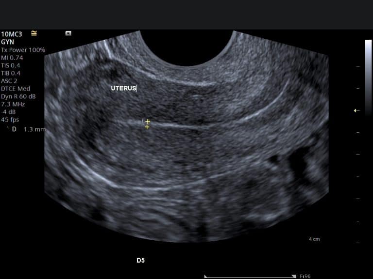
An internal ultrasound allows our ultrasonographer to look through the vagina. During the procedure, you will be asked to lie on your back and a small probe will be gently passed into the vagina and the images are transmitted to a monitor.
Internal examinations are ideal for looking at your ovaries and womb and may cause some discomfort, but should not be painful.
Transrectal Ultrasound
A transrectal ultrasound allows our ultrasonographer to look through the rectum (back passage). During the procedure, you will be asked to lie on your side and a small probe will be gently passed into your rectum and the images are transmitted to a monitor.
Transrectal examinations are ideal for looking at the prostate or when a transvaginal scan is not possible, for example in patients with virgo intacta.
Preparing for an Ultrasound Scan
General Preparation Guidelines
While the specifics can vary based on the type of ultrasound, some general guidelines are universally applicable.
- Fasting: For certain types of ultrasounds, like those examining the abdominal area, you might need to fast for several hours beforehand. As a study from the University of California, San Francisco, notes, fasting helps to clear the stomach and intestines, providing clearer images.
- Hydration: For some scans, particularly pelvic ultrasounds, being well-hydrated is crucial. A study published in the American Journal of Roentgenology suggests that a full bladder can help improve the visibility of pelvic organs.
- Clothing: Wear comfortable, loose-fitting clothing to your appointment. You may be asked to change into a gown, but comfortable clothes make the process easier before and after the scan.
Specific Preparation for Different Types of Ultrasound
- Abdominal Ultrasound: If you’re having an abdominal ultrasound, as mentioned earlier, fasting is usually required. The University of Cambridge recommends fasting for at least 6 hours before your scan to ensure the most accurate results.
- Pelvic Ultrasound: For a pelvic ultrasound, drinking water before your scan and not urinating can help. Studies have shown that a full bladder pushes intestinal gas out of the pelvis, allowing for better imaging.
- Musculoskeletal Ultrasound: No special preparation is typically needed for musculoskeletal ultrasounds. However, wearing clothing that allows easy access to the area being examined is advisable.
Interpreting Ultrasound Results
Understanding the results of an ultrasound scan is a crucial aspect of the diagnostic process. Ultrasound, a widely used medical imaging technique, provides real-time, detailed images of the body’s internal structures. Its interpretation, however, requires professional expertise.
Understanding the Images
Ultrasound images are created using high-frequency sound waves, which bounce off tissues, organs, and fluids in the body. These echoes are captured and transformed into visual images. A study from the American College of Radiology highlights that interpreting these images requires an understanding of various factors, such as the density of the tissues and the angle of the ultrasound probe.
Professional Expertise
Interpretation of ultrasound results is typically done by a radiologist or a trained sonographer. They analyze the shapes, sizes, and textures of structures and organs visible in the scan. As noted in a study published in the British Journal of Radiology, the expertise of the interpreter plays a significant role in the accuracy of the diagnosis.
Types of Findings
Ultrasound scans can reveal a range of findings. For example, in obstetrics, ultrasound is essential for monitoring fetal development, as shown in research detailed in the Journal of Obstetrics and Gynecology. In other areas, such as cardiology or abdominal imaging, ultrasound helps in identifying abnormalities like tumors, cysts, or fluid buildup.
Real-Time Observation
One of the significant advantages of ultrasound, as discussed in a study published in the European Journal of Radiology, is its ability to provide real-time observations. This aspect is particularly useful in procedures like needle biopsies, where the ultrasound guide ensures precision.
Normal vs. Abnormal Findings
The interpretation of an ultrasound also involves distinguishing between normal and abnormal findings. For instance, a study in the Journal of Clinical Ultrasound demonstrates how sonographers differentiate between healthy and diseased tissues based on their appearance in the ultrasound images.
Communication of Results
After the ultrasound examination, the interpreting physician prepares a report detailing the findings. As emphasized in research by the Radiological Society of North America, this report plays a critical role in guiding further medical management or treatment.
Importance of Follow-Up
In some cases, additional tests may be recommended. A study conducted at Harvard Medical School entitled “Ultrasound Diagnostics: Beyond the Image” showed that follow-up tests like CT scans, MRIs, or biopsies might be necessary to further investigate certain findings.
Patient Involvement
Finally, it is important for patients to discuss their ultrasound results with their healthcare provider. A study published in the Annals of Family Medicine suggests that patient understanding of ultrasound results can significantly impact treatment decisions and patient compliance.
Interpreting ultrasound results is a complex process that combines technology, expertise, and clinical knowledge. It’s not just about the images themselves, but about understanding what they mean in the broader context of a patient’s health and medical history.
Safety and Efficacy of Ultrasound Scanning
Ultrasound scanning, a cornerstone of modern diagnostic imaging, stands out for its safety and efficacy. Its ability to provide real-time visualizations of internal bodily structures without the use of ionizing radiation makes it a preferred choice in various medical scenarios.
Safety: A Non-invasive and Radiation-free Technique
One of the most significant advantages of ultrasound scanning is its non-invasive nature. Unlike other imaging modalities, such as X-rays or CT scans, ultrasound does not use ionizing radiation. This aspect is crucial, especially in sensitive populations like pregnant women and children. As G. ter Haar noted in their research published in Interface Focus, ultrasound imaging is safe and free from the risks associated with radiation exposure, making it suitable for routine and repeated use in monitoring pregnancies and diagnosing various conditions.
Efficacy: Accuracy and Versatility in Diagnosis
The efficacy of ultrasound scanning lies in its versatility and accuracy. Ultrasound can be used to examine various parts of the body, including the abdomen, heart, blood vessels, and musculoskeletal system. According to a study in the Journal of Ultrasound in Medicine by M. Blaivas, ultrasound has proven highly effective in the evaluation of pneumonia, rivaling even CT scans in accuracy for certain conditions. This highlights the technology’s ability to provide detailed and accurate images, essential for correct diagnosis and treatment planning.
Real-Time Imaging for Immediate Assessment
A unique feature of ultrasound scanning is its ability to provide real-time images. This capability allows healthcare professionals to conduct immediate assessments and make timely decisions, especially in emergency settings. As indicated in a study published in Clinical Radiology by D. Lipscomb et al., ultrasound’s real-time imaging has been instrumental in diagnosing and managing pleural diseases, showcasing its critical role in both diagnosis and procedural guidance.
Patient Comfort and Convenience
Ultrasound scans are generally quick and painless, factors that significantly enhance patient comfort. The non-invasive nature of the procedure means that it is usually well-tolerated by patients of all ages. This aspect of patient comfort and convenience is emphasized in studies like the one by Juha Havukumpu and colleagues, where the ease of use and painless nature of ultrasound scanning are highlighted.
Ongoing Advances in Ultrasound Technology
Continual advancements in ultrasound technology further augment its safety and efficacy. Innovations such as 3D/4D imaging, as discussed by T. Whittingham, have opened new avenues for more detailed and informative scans. These advancements not only enhance the diagnostic capabilities of ultrasound but also contribute to more effective patient management and treatment outcomes.
Ultrasound scanning, therefore, stands as a testament to the progress in medical imaging, offering a safe, effective, and patient-friendly diagnostic tool. Its role in modern healthcare continues to expand, driven by technological advancements and its inherent advantages.
Where to get an ultrasound scan in London?
If you are looking for a private ultrasound in London to reduce the NHS waiting times and get quick answers for your health, you can rest assured that our sonographers have the skills and the knowledge to provide you with the instant answers you need. At International Ultrasound Services we offer diagnostic ultrasounds that can help to reassure you and in cases of any abnormalities to seek medical help. You can now book your appointment online or over the phone.
The private scans are performed by highly qualified sonographers who perform precise examinations to help predict and diagnose any situation that might arise. Our patients benefit from early diagnosis and treatment.
Ultrasound scanning stands as a cornerstone in contemporary medical diagnostics, offering a window into the human body with unparalleled safety and precision. Its versatility extends across various medical fields, from monitoring the intricate development of a fetus in obstetrics to aiding in the precise diagnosis of musculoskeletal conditions. A study from the Journal of Ultrasound in Medicine highlighted how ultrasound has revolutionized the evaluation of pneumonia, underscoring its significance in critical care and emergency medicine.
The technology’s non-invasive nature, highlighted by research in Interface Focus, makes it a patient-friendly option, devoid of the risks associated with ionizing radiation. This aspect is crucial, considering the increasing use of ultrasound in routine health checks and specialized diagnostics. Further advancements, as discussed in Progress in Biophysics and Molecular Biology, have led to innovations like 3D and 4D imaging, enhancing the depth and breadth of ultrasound’s diagnostic capabilities.
The real-time aspect of ultrasound imaging, as explored in a study published in the Journal of Digital Imaging, allows for immediate insights into the body’s internal structures, facilitating prompt and effective medical decision-making. This immediacy is invaluable in fast-paced medical environments and contributes significantly to patient care.
As ultrasound technology continues to evolve, its applications are likely to expand even further, making it an indispensable tool in modern healthcare. Its ability to provide detailed, real-time images safely and efficiently ensures that ultrasound scanning remains at the forefront of diagnostic imaging, offering peace of mind and critical information to both healthcare providers and patients alike.
Do you get ultrasound results immediately?
If you had your scan in the NHS, it is more likely that you will have to wait a few days until the report reaches your GP or consultant. If you had your scan in a private clinic you will probably receive your results immediately or within a few hours.
What is a Doppler ultrasound scan?
Colour Doppler or colour flow Doppler, which was discovered by Christian Doppler in 1841, is the technology to visualize blood flow during an ultrasound scan. Colour flow is being used all the time these days as it helps to differentiate between different structures, for example, a blood vessel and a cyst that can have a similar appearance on ultrasound. It helps to evaluate the vascularity of a tumour found for example in the breast scan, pelvic scan or liver scan. It helps to evaluate the vascularity of whole organs such as in the thyroid scan in cases of thyroiditis.
Do you need a private ultrasound in London?
How accurate is an ultrasound scan
Ultrasound scans are accurate in diagnosing specific diseases. Accuracy however depended on the experience of the sonographer or radiologists as well as the quality of the ultrasound scanner.
How much is an ultrasound scan?
There various factors affecting the price you pay for an ultrasound scan, such as the location and experience of the operator. You can find the prices of our ultrasound scans is clearly marked on the specific examination, or you see all the prices of ultrasounds.
FAQs about Ultrasound
What is an Ultrasound Scan?
An ultrasound scan, also known as sonography, utilizes high-frequency sound waves to create images of the inside of the body. This technology is widely used due to its non-invasive nature and does not involve exposure to radiation. Studies have shown ultrasound to be a safe and effective diagnostic tool in various medical fields (Havukumpu et al., 2008).
How Should I Prepare for an Ultrasound?
Preparation for an ultrasound can vary depending on the type being performed. For example, you may need to fast for a few hours before an abdominal ultrasound. It’s important to follow the specific instructions provided by your healthcare provider to ensure accurate results.
Is Ultrasound Scanning Painful?
Ultrasound scanning is generally painless. As Juha Havukumpu and colleagues (2008) noted, it’s a non-invasive procedure typically involving the application of a small amount of gel on the skin and moving a handheld probe over the area of interest.
How Long Does an Ultrasound Take?
The duration of an ultrasound scan can vary. Most scans are completed within 30 minutes to an hour. The specific time depends on the area being examined and the complexity of the scan.
Can Ultrasound Detect Cancer?
Ultrasound is a valuable tool in detecting and monitoring various conditions, including cancer. It helps in visualizing tumors and can be pivotal in guiding biopsies and other diagnostic procedures.
Are There Any Risks Associated with Ultrasound Scans?
Ultrasound is considered to be a very safe diagnostic tool. As noted by G. ter Haar (2011), the lack of ionizing radiation makes it safer compared to other imaging methods like X-rays.
Can I Get an Ultrasound Scan During Pregnancy?
Yes, ultrasound scans are a routine part of prenatal care. They are used to monitor the health and development of the fetus throughout pregnancy.
What Can I Expect After an Ultrasound Scan?
Typically, there are no special precautions to take after an ultrasound scan. You can usually return to your normal activities immediately unless advised otherwise by your healthcare provider.
How Accurate are Ultrasound Scans?
Ultrasound scans are a highly accurate diagnostic tool when performed by skilled technicians. The accuracy can depend on various factors, including the type of scan and the specific condition being assessed.
Can Ultrasound Help in Treatment Planning?
Absolutely. Ultrasound provides real-time images that are crucial in planning and guiding various treatments and surgical procedures.
Private Ultrasound
If you are looking for reassurance early in your pregnancy (not 4D ultrasound) or a general medical private ultrasound in London to visualise the internal organs and body structures to get quick answers about your health, you can rest assured that our sonographers have the skills and the knowledge to provide you with the instant answers you need.
References:
NHS – Ultrasound Scan
FDA – Ultrasound Imaging
NHS- Ultrasound Scans in Pregnancy
NHS – 12-week scan
ROOG – Guidance for antenatal screening and ultrasound in pregnancy in the evolving coronavirus pandemic
NICE – Antenatal care for uncomplicated pregnancies
BMUS – Guidelines for the safe use of diagnostic ultrasound equipment
Ultrasound in Obstetrics and Gynaecology – ISUOG Practice Guidelines: performance of first‐trimester fetal ultrasound scan
Healthline – Pregnancy Ultrasound
WHO – WHO recommendation on early ultrasound in pregnancy
Medically Reviewed by Tareq Ismail Pg(Dip), BSc (Hons)
[post-views]
About the Author: Yianni Kiromitis PgC (Medical Ultrasound), BSc(Hon)
I have 15+ years in ultrasound scanning and more than 20 years experience in Diagnostic Imaging. I have worked for multiple NHS hospitals as well as in the private sector.


