Early detection saves lives, especially when it comes to breast health and the potential risks of breast cancer.
Breast ultrasound is a powerful tool that can help detect breast abnormalities and bring them to a doctor's attention quickly.
In this article, we will take a look at the importance of early detection in breast health and how breast ultrasound can play a role in it.
Overview of Breast Health
Breast health is an important factor in maintaining overall wellness for women. To maximize this, regular check-ups and diagnostic services are important. Early detection of abnormalities can lead to quick and successful treatment that can improve outcomes and save lives.
Thanks to advances in technology, there are now several types of imaging screenings available that can help detect breast cancer and other abnormalities in the breasts. One such type of imaging is breast ultrasound, which uses high-frequency sound waves to create a detailed image of the breasts’ internal structures. This type of imaging has some advantages over other forms like mammography as it tends to be more suitable for younger women, as well as those with dense breast tissue or implants.
There are two types of ultrasound machines that can be used to diagnose breast health issues: standard ultrasound (also known as B-mode) and Doppler ultrasound (also known as Duplex). Both use ultrasound waves and a transducer that sends pulses into the patient’s body. These pulses generate pictures on a computer monitor showing results from inside the body which are then used by medical professionals to analyze any potential issues or abnormalities within the breasts.
Early detection has been proven time again to be one most reliable methods for successfully managing or treating ailments such as breast cancer, or diagnosing benign conditions like fibrocystic changes or fibroadenomas that can potentially lead up to cancer if left untreated for long periods of time. As such, regular check-ups should become part of everyone's prevention plan when it comes to maintaining their breast health, making use of all diagnostic tools available from conventional screenings such as mammography to individually tailored ones like Breast Ultrasound when needed.
Risk Factors for Breast Cancer
The development of breast cancer is complex, and it is difficult to predict who will develop the condition. However, several risk factors have been identified that may increase the chance of developing it. The risk can be categorized as modifiable (e.g., lifestyle) or non-modifiable (e.g., family history), and this can help patients better understand their risks and possible ways to reduce them.
The most commonly discussed modifiable risk factor associated with breast cancer is an individual’s lifestyle. Specifically, individuals who consume large amounts of alcoholic beverages, engage in physical inactivity, are overweight or obese, or underwent hormone replacement therapy (HRT) or oral contraceptive use may experience an increased risk for breast cancer compared to those who do not engage in these behaviors. Other possible factors include exposure to certain environmental hazards such as radiation or various occupational chemical exposures.
Beyond lifestyle factors, there are numerous non-modifiable risk factors for developing breast cancer, including a strong family history of the condition, age at menarche (earlier onset maturation), age at first childbirth (later onset motherhood), nulliparity (never having given birth), personal history of benign breast disease (BBD), genetic markers such as BRCA mutations and more recently DNA methylation patterns around gene promoters have been seen to influence the onset of BC as well Lastly, race and ethnicity should be accounted for when discussing personal risks associated with this particular health condition.
Breast Ultrasound
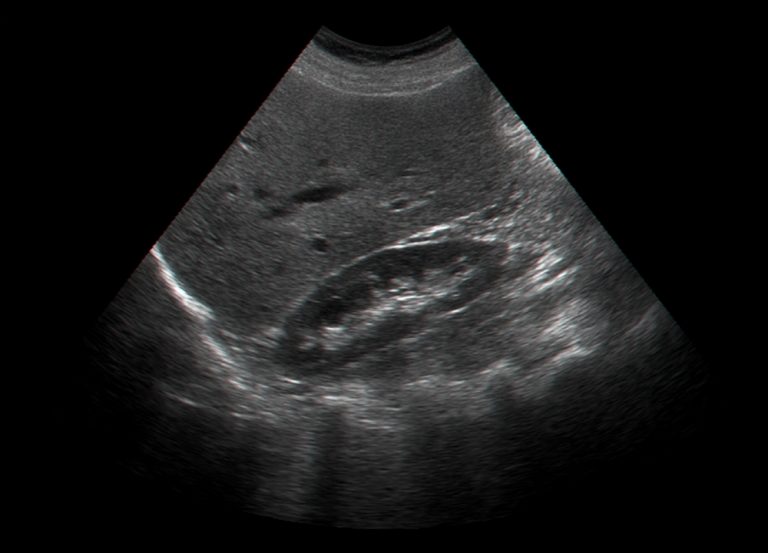
Breast ultrasound is an imaging technique that utilizes high frequency sound waves to create a picture of the breast tissue. It has proven to be an effective diagnostic tool that can help detect the early stages of breast disease and cancer.
This article will cover the basics of breas ultrasound and its importance in detecting breast issues.
What is a Breast Ultrasound?
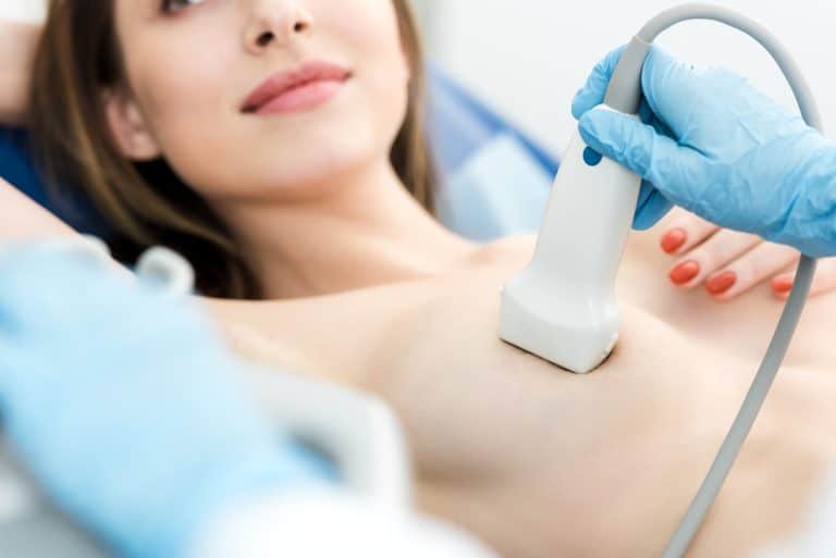
Breast ultrasound is a medical imaging technique used to visualize the internal structures of the breast. This imaging procedure is non-invasive, which means it does not involve radiation or require any type of anesthesia. During a breast ultrasound, sound waves are used to create an image of the breast tissue. The interpretation of these images can help detect any potential problems that may be present in the breasts and enable early diagnosis and treatment.
Breast ultrasound can detect abnormalities in the size, shape, texture, and density of the breast tissue. It can also detect changes in breast density due to age, hormone imbalances or certain medications; as well as lumps or tumors. In some cases, breast ultrasounds may be used along with other imaging techniques such as mammography to ensure accurate diagnosis of potential issues within the breasts.
Ultrasound technology has come a long way in recent years due to technological advancements that allow for more accurate images at significantly lower exposures than ever before. Ultrasound images are now able to provide detailed information about even very small lesions and suspicious areas within the breasts; allowing healthcare providers to give patients timely diagnoses and personalized treatments for better outcomes and earlier detection of serious conditions such as cancer.
Benefits of Breast Ultrasound
Breast Ultrasound is a fast and reliable imaging technology used to detect problems such as breast lumps or cancer. It is also useful in detecting early signs of breast disease that may not be detected through a standard X-ray (mammogram). Ultrasound can provide women with peace of mind that their breasts are healthy, and it can also give doctors additional information to aid in diagnosis and treatment decisions.
The benefits of using this technology for early detection include:
- Lower radiation exposure compared to X-ray mammography
- No compression is needed, unlike mammograms which require compressing the breast
- Images are easier to interpret due to higher contrast due to ultrasound’s non-ionizing radiation
- Ability to image cysts, which usually appear more clearly while not as visible with X-rays
- Avoids unnecessary biopsies
- Can be used as an adjunct screening method along with conventional mammography when lumps or abnormalities are detected
Early Detection
Early detection of breast cancer is important for the most successful treatment. Mammograms, while being an important tool, can lead to false positives in some cases. This is where a breast ultrasound can play a crucial role in assisting with early detection.
This can give a more precise diagnosis, allowing doctors to diagnose a condition and treat it in the early stages. Knowing the benefits of breast ultrasound can help you decide if it’s right for you.
Early Detection and Breast Cancer Treatment
The impact of early detection on breast cancer cannot be overstated. According to the American Cancer Society, when breast cancer is detected in its early stages, it is easier to treat and generally has a better prognosis. Additionally, earlier diagnosis makes it more likely that any necessary treatment will require minimal intervention.
One effective way to detect the presence of breast cancer in its earliest stages is through ultrasound imaging. Breast ultrasound is a safe and non-invasive imaging technique that uses sound waves to create pictures of organ structures or internal soft tissue within the body. It allows practitioners to visualize abnormal masses that can’t be seen with other traditional imaging techniques and can be used to detect both benign and malignant tumors.
Breast ultrasound can help identify abnormalities by focusing on specific areas inside the breasts where suspicious signs indicate possible treatments or monitoring may be necessary - even if no signs are visible during physical examination or mammography screening, making it an incredibly important tool in this fight against breast cancer. By identifying potential problems before they become serious issues, early detection through breast ultrasounds makes successful treatment much more likely. This procedure is also non-ionizing radiation free meaning patients need not worry about any additional exposure during the exam besides sound waves which are completely harmless when used as intended for medical diagnosis purposes.
Early Detection and Breast Cancer Survival Rates
Timely detection of breast cancer is critical to increasing one’s chances of surviving the disease. Early detection is key in maximizing the chances of effective treatment. By performing regular self-exams and participating in mammograms, physician-referred screenings, and ultrasound screenings when necessary, patients can increase their odds of detection at an early stage.
Mammograms are a key screening tool in early breast cancer detection and typically recommended for women 40 years and older on an annual basis. However, mammograms cannot detect all types of breast cancer. To screen for all known forms of breast cancer confidently and accurately, Breast Ultrasound can be used as an effective complement to the standard mammogram procedure.
Ultrasound can help detect lumps or masses in dense or hard-to-image regions within the breasts that may not be immediately visible on a mammogram or other imaging exams such as MRI (Magnetic Resonance Imaging). It is also accurate at detecting very small lesions while minimizing false positives (identifying something as abnormal when it isn’t). Early infantile diagnosis remains vital and 3D ultrasounds allow physicians to measure and analyze developing features in newborns more effectively than ever before.
Studies show that when compared with people whose cancers were diagnosed at later stages, those individuals whose cancers were detected early have greater survival rates. According to the National Cancer Institute, “If every woman over age 50 had her own yearly mammogram screening beginning between 1990 and 1995, approximately 32% fewer deaths from breast cancer would have occurred over a six-year period for women age 55 to 69." As such, early detection continues to be essential for both reducing mortality from this often deadly form of malignancy and heightening successful treatment outcomes.
Conclusion
In conclusion, early detection is key in the prevention and treatment of breast health issues. Breast ultrasound is an invaluable tool in this process, as it allows for the detection of potential issues and abnormalities that may not be visible from a mammogram. It is highly recommended that women of all ages conduct regular breast ultrasound screening in order to ensure the highest level of breast health.
The Importance of Early Detection
The importance of early detection in breast health cannot be overstated. Unfortunately, many women fail to get the medical care they need most until cancer is advanced and treatment becomes more difficult. In less advanced stages, some forms of breast cancer have a five-year survival rate of greater than 95%. Unfortunately, because screenings can be expensive or lack availability in some rural areas, less than 50% of women with breast cancer are diagnosed with an early stage disease. Therefore, it is critical for all women to actively seek early detection resources to improve their chances for successful outcomes.
Breast ultrasound is one tool that can be used to help detect changes in the breasts that may require further evaluation or biopsy to determine if a woman has cancerous cells. This test complements mammography as both are important tests when it comes to detecting any changes in the breasts. When performed by trained personnel, ultrasound is capable of revealing issues such as fibroadenomas and cysts that can occur from time-to-time and neither of these are typically visible or detectable through traditional mammography techniques.
Although not all detected changes will lead to biopsy confirmation for cancer, women need to be aware that early detection will save them valuable time and money for other tests such as MRI scans or further biopsies if the basic testing does not produce a harder diagnosis.
How Breast Ultrasound Can Help
Breast ultrasound is an excellent tool for doctors to use to gain a better picture of the breast tissue, which is helpful in identifying any abnormalities. This imaging technique can detect changes in breast tissue as small as 2mm and can detect lumpy areas or masses. The size and shape of the mass can be accurately determined with a 3-dimensional ultrasound image, providing a more accurate diagnosis of the abnormality.
Not only does ultrasound allow doctors to diagnose a possible problem, but it also gives them the opportunity to evaluate a change over time if they suspect that something may be developing. By analyzing the composition of the mass and by measuring any changes made over time, doctors are able to take appropriate steps—such as biopsy—if necessary.
Breast ultrasound can also be used as a screening tool for patients who may have previously had growths that have been removed or for women who have family histories of certain types of cancer and higher risk factors for cancerous tumors in their breasts. It is especially useful for patients getting mammograms because women with dense breasts are more likely to have false positives on their mammograms, making it difficult for radiologists to make an accurate diagnosis without further examination.
In addition, breast ultrasound aids physicians in making decisions about treatment options due to its ability to provide detailed information about many different components within the breast tissue at various levels. Ultrasound allows doctors to see almost immediately any suspicious changes or lesions which will enable them to plan effective treatment as soon as possible in cases that require intervention and management of care when needed.
Content Information
We review all clinical content annually to ensure accuracy. If you notice any outdated information, please contact us at info@iuslondon.co.uk.
About the Author:

Dr. Mohammad Jalaleddin (GMC number: 7729988) is an internationally-trained physician who earned his medical degree from Tishreen University in Syria in 2005. He completed his specialization in diagnostic radiology in 2009, followed by a Master's degree in 2011, which focused on the radiological alterations of bone grafts. Dr. Jalaleddin has worked as a radiologist in various hospitals and clinics, developing significant expertise in ultrasound and MRI scans. He has extensive experience in performing a wide range of ultrasound examinations, including breast, abdominal, pelvic, and vascular studies. In 2017, he moved to the UK and completed the requalification process with the General Medical Council (GMC). Since March 2021, he has been a part of the Imperial College Healthcare NHS Trust. Dr. Jalaleddin is a member of the General Medical Council (GMC) and the British Medical Ultrasound Society (BMUS).


