If you've recently had an abdominal ultrasound, you might be finding the medical terminology in the report challenging to understand.
What does an abdominal ultrasound report cover?
Typically, an abdominal ultrasound report will include details on key organs within the abdomen, such as the liver, gallbladder, pancreas, kidneys, and spleen. Each of these organs is assessed for size, structure, and any abnormalities.
Most abdominal ultrasound reports don’t cover pelvic organs, as these are typically included in a separate pelvic ultrasound. If you’ve had both an abdominal and pelvic scan, you can combine the insights here with those in the "Understanding Your Pelvic Ultrasound Report" article for a complete picture.

Ultrasound Terminology for Each Abdominal Organ
To help clarify the ultrasound terminology used, we will go over some key terms by organ, starting with the liver.
The Liver
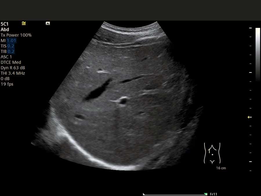
In ultrasound reports, one of the main elements described is the liver's echotexture, which refers to the appearance and texture of liver tissue on the scan.
The liver can have:
- Normal Echotexture: A smooth, uniform appearance indicating healthy liver tissue.
- Heterogeneous Echotexture: This term describes a patchy or uneven appearance, often caused by conditions such as fatty liver, hepatitis, cysts, or masses.
- Echogenic or Hyperechoic: The liver appears brighter than normal, commonly associated with fatty liver disease or hepatic steatosis.
- Hypoechoic: A darker appearance than expected, though this is rare. In over 20 years of ultrasound experience, I’ve rarely observed hypoechoic livers.
- Coarse Echotexture: This describes a rougher, less uniform appearance of liver tissue and often indicates chronic liver disease.
Additionally, the appearance of the liver capsule (the outer layer of the liver) is also evaluated. A smooth capsule is typical in a healthy liver, while a nodular or irregular capsule may indicate fibrosis or cirrhosis and is often seen alongside coarse liver parenchyma.

Portal Vein
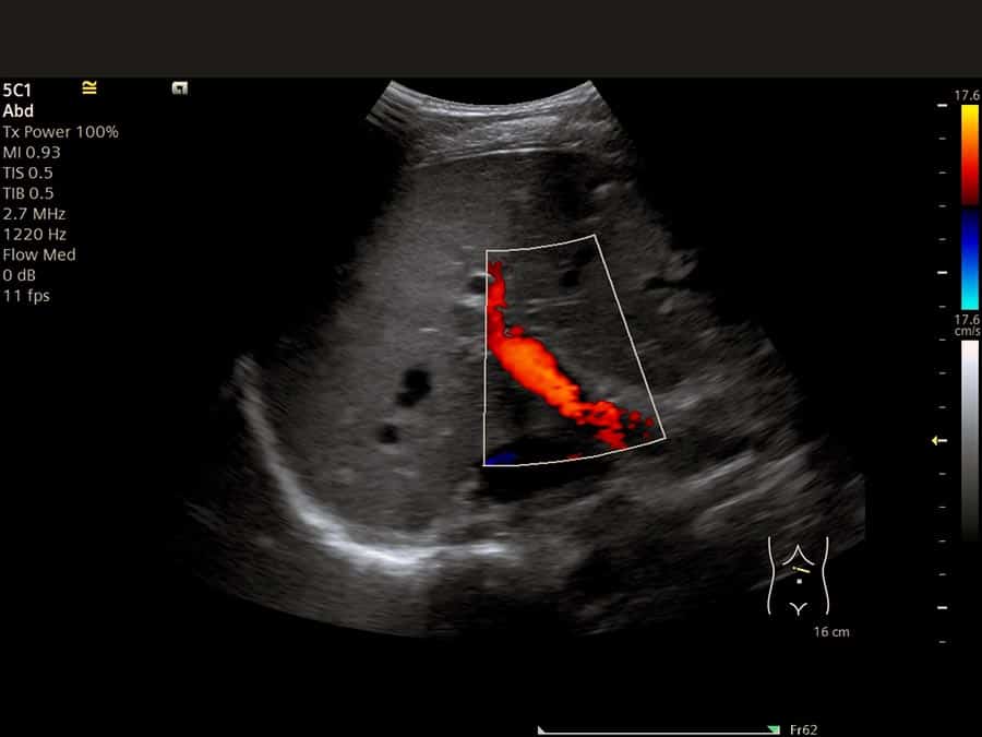
The portal vein serves as the primary source of blood supply to the liver, delivering nutrient-rich blood absorbed from the intestines. In an ultrasound report, the flow direction in the portal vein is typically noted, which can indicate:
- Hepatopetal flow (flow toward the liver): Normal direction.
- Hepatofugal flow (flow away from the liver): Often associated with advanced liver cirrhosis.
- Absent flow: May indicate a blockage or clot in the portal vein, necessitating further imaging for confirmation.
Hepatic Veins
Blood enters the liver through the portal vein and exits via the hepatic veins. These veins are often not detailed in ultrasound reports because they are harder to visualize and are usually found to be normal.
Gallbladder
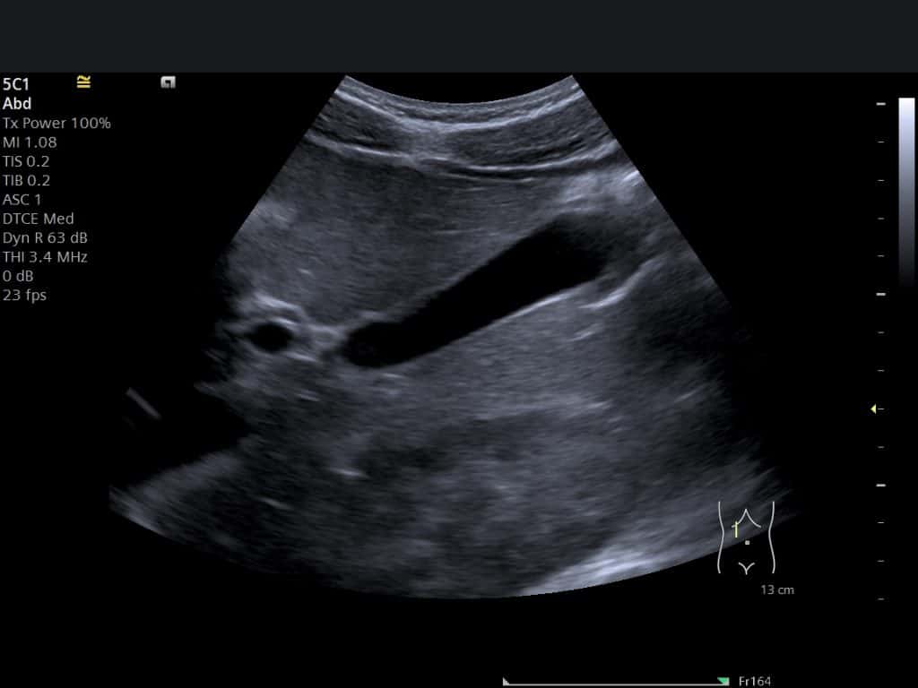
The gallbladder stores bile, and gallstones are a common cause of abdominal pain. In an ultrasound report, the gallbladder is assessed for any abnormalities, including wall thickening, the presence of stones, or bile sludge. These details help evaluate gallbladder health and potential issues causing symptoms.
Common Bile Duct (CBD)
The common bile duct is a channel that connects the gallbladder to the intestine, allowing bile to flow into the digestive tract. Ultrasound reports will either confirm if the bile duct appears normal or specify its diameter.
The normal diameter of the bile duct varies depending on the patient’s age and whether the gallbladder is present or removed (due to cholecystectomy). A widened or "dilated" bile duct can indicate an obstruction, such as a stone within the duct or a mass pressing on and blocking it.


The Pancreas
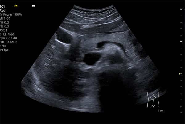
The pancreas is located in the central upper abdomen, but it can be challenging to visualize clearly on an ultrasound. This difficulty arises because loops of bowel often lie between the pancreas and the ultrasound probe, obstructing a clear view. As a result, it’s common for ultrasound reports to note that the pancreas is partially visualized, with only the head and part of the body typically visible.
Kidneys
Ultrasound evaluation of the kidneys assesses both their size and echotexture. When the renal parenchyma appears unusually bright (echogenic), it may signal diffuse renal disease. Other common kidney conditions detectable by ultrasound include hydronephrosis, renal cysts, kidney stones, and, in some cases, tumors.
Spleen
The spleen, located just above the left kidney, is also evaluated for its size and echotexture during an ultrasound scan. An enlarged spleen may suggest underlying liver disease or other systemic conditions.
You can find more information about the ultrasound terms used in your report in our ultrasound glossary.

Content Information
We review all clinical content annually to ensure accuracy. If you notice any outdated information, please contact us at info@iuslondon.co.uk.
About the Author:

Yianni is a highly experienced sonographer with over 21 years in diagnostic imaging. He holds a Postgraduate Certificate in Medical Ultrasound from London South Bank University and is registered with the Health and Care Professions Council (HCPC: RA38415). Currently working at Barts Health NHS Trust, Yianni specialises in abdominal, gynaecological, and obstetric ultrasound. He is a member of the British Medical Ultrasound Society (BMUS), Society of Radiographers (SoR) and regularly contributes to sonographer and junior radiologists training programs.


