Pelvic Infections or Pelvic Inflammatory Disease (PID), is a term used for infection of Pelvic organs i.e the Uterus, Fallopian tubes & Ovaries in a woman. These are commonly transmitted sexually but may, sometimes, be attributed to other causes. According to WHO, about 448 million new cases of Sexually Transmitted Infections (STI) are diagnosed annually and it is among the top 5 disease categories for which an individual seeks medical care.
Signs and Symptoms – Many women having PID may not have any obvious symptoms, but usually an episode of PID or Pelvic infections may present with the following symptoms:
- Lower abdominal pain
- Pain during intercourse
- Bleeding after intercourse
- Irregular/ abnormal periods or spotting in between two periods.
- Excessive or foul-smelling vaginal discharge
- Vaginal or Perineal itching
- Frequent or painful/burning urination
- Occasionally, in advanced cases, there may be fever, vomiting, severe pain or even fainting episodes.
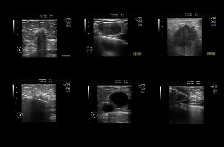
Pelvic ultrasound is the first line of investigation for suspected pelvic inflammatory disease. You can find more about the investigation at our pelvic ultrasound page.
Complications – Pelvic infections and PID can be a cause of significant morbidity and may have long-lasting outcomes including:
- It is the leading cause of infertility, about 1 in 8 women having a history of PID can have difficulty in getting pregnant.
- It may lead to chronic pelvic pain in about 25% of women. They may have pain related to menstrual cycles or may have persistent lower abdominal pain.
- PID leads to the formation of adhesions i.e. scar tissue in or around fallopian tubes which significantly increases the chances of Ectopic pregnancy (pregnancy implanted outside the cavity of the uterus that can lead to serious life-threatening complications)
- In the long-term, recurrent pelvic infection, especially with HPV, can be a precursor of cervical cancer.
Causes and Risk factors – PID is generally considered to be a polymicrobial infection, i.e it is caused by multiple micro-organisms. These generally include bacterial pathogens like Chlamydia and Neisseria along with a number of other pathogens like Gardnerella, Mycoplasma, Trichomonas, Herpes Simplex Virus-2 and various anaerobic bacteria that may be transmitted by sexual contact and are found in the vagina. Hence, it comes as no surprise that PID results primarily from unprotected sexual intercourse in most cases. However, there may be other causes for the development of the infection and the following factors may increase the risk of a woman suffering from PID:
- Unprotected sexual activity i.e. intercourse without using a condom.
- Having multiple sexual partners or having intercourse with a person who has multiple sexual partners.
- The onset of sexual activity before the age of 25 years.
- A history of the prior sexually transmitted disease which has been incompletely treated in the woman or the sexual partner.
- A history of sexual abuse
- Any history of Gynaecological interventional procedure for e.g. Endometrial biopsy, IUCD insertion, Hysteroscopy etc.
- Vaginal douching has been paradoxically associated with the development of a vaginal infection as it alters the normal vaginal balance of useful versus harmful bacteria. However, some studies have failed to demonstrate a clear association between the two.
- Apart from these, certain genetic factors have been studied which are found to predispose to pelvic infection.
- Any decrease in generalized body immunity may also cause a flare-up of an underlying infection for e.g. – in prolonged illness, HIV infection or any immune-compromised state such as pregnancy.
Pelvic Infection Treatment and Prevention – PID or Pelvic Infection treatment is usually by an antibiotic course along with other medications lasting for about 2 weeks. Depending upon your symptoms, this may be either an oral medication or sometimes, in severe cases, a woman may need to be hospitalised for injectable medications or surgical intervention as required. It is important for the sexual partner to be treated simultaneously to prevent re-infection.
However, PID and sexually transmitted infections are better prevented than treated. Hence, anyone who is at risk of pelvic infections should take the following precautions:
- Practice safe sex i.e. always use a condom at the time of intercourse (unless of course, you are actually trying for pregnancy)
- Avoid indulging in indiscriminate sexual activity with multiple partners or with a partner who is in a sexual relationship with multiple persons.
- Avoid indulging in sexual activity at a very young age.
- Consult a doctor at the first sign of infection & take proper treatment.
- Practice good perineal hygiene and avoid vaginal douching. It helps to wipe from the front backwards after passing urine/stools rather than wiping from back to front.
- Consume a variety of fruits, probiotics and a healthy, well-balanced diet to boost your immunity.
Content Information
We review all clinical content annually to ensure accuracy. If you notice any outdated information, please contact us at info@iuslondon.co.uk.
About the Author:

Yianni is a highly experienced sonographer with over 21 years in diagnostic imaging. He holds a Postgraduate Certificate in Medical Ultrasound from London South Bank University and is registered with the Health and Care Professions Council (HCPC: RA38415). Currently working at Barts Health NHS Trust, Yianni specialises in abdominal, gynaecological, and obstetric ultrasound. He is a member of the British Medical Ultrasound Society (BMUS), Society of Radiographers (SoR) and regularly contributes to sonographer and junior radiologists training programs.


