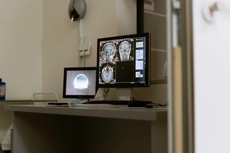
Medical diagnosis forms the foundation of effective healthcare. It's the process through which healthcare professionals identify a medical condition or disease based on a patient's signs, symptoms, and test results. Accurate diagnosis is critical for determining the most appropriate treatment plan and ensuring optimal patient outcomes.
Over the years, diagnostic techniques have evolved significantly, incorporating advanced technologies to improve accuracy and efficiency. Among these technologies, ultrasound has emerged as a powerful tool in the diagnostic arsenal of healthcare providers.
Evolution of Diagnostic Techniques
Historically, medical diagnosis relied heavily on physical examinations and patient histories. Doctors would use their senses - sight, sound, touch, and sometimes even smell - to detect abnormalities and make educated guesses about a patient's condition. While these methods remain valuable, they have limitations in detecting internal issues or subtle changes within the body.
The advent of imaging technologies revolutionized medical diagnosis. X-rays, discovered in 1895, were the first to allow doctors to see inside the human body without surgical intervention. This breakthrough paved the way for more advanced imaging techniques, including computed tomography (CT), magnetic resonance imaging (MRI), and ultrasound.
Ultrasound: A Key Player in Modern Diagnostics
Ultrasound technology, also known as sonography, uses high-frequency sound waves to create real-time images of structures within the body. The basic principle involves sending sound waves into the body and analyzing the echoes that bounce back.
This imaging modality offers several advantages:
- Non-invasive: Ultrasound doesn't require any incisions or injections.
- Real-time imaging: It provides immediate, moving images of internal structures.
- No radiation: Unlike X-rays or CT scans, ultrasound doesn't use ionizing radiation.
- Cost-effective: Compared to other imaging techniques, ultrasound is relatively inexpensive.
- Portable: Many ultrasound machines are compact and can be moved easily between rooms or even used in the field.
These benefits have made ultrasound a go-to diagnostic tool in many medical specialties.
Common Applications of Ultrasound in Diagnosis
Obstetrics and Gynecology
Ultrasound has become indispensable in prenatal care. It allows healthcare providers to:
- Confirm pregnancy and estimate gestational age
- Assess fetal development and growth
- Detect multiple pregnancies
- Identify potential complications or abnormalities
- Guide procedures such as amniocentesis
Beyond pregnancy, ultrasound is used to examine the uterus, ovaries, and other pelvic structures, aiding in the diagnosis of conditions like ovarian cysts, fibroids, and certain cancers.
Cardiovascular Health
Echocardiography, a specialized form of ultrasound, is used to examine the heart's structure and function. It can help diagnose:
- Heart valve problems
- Congenital heart defects
- Cardiomyopathies (diseases of the heart muscle)
- Blood clots or tumors in the heart
Vascular ultrasound is used to assess blood flow in arteries and veins, helping to detect conditions like deep vein thrombosis, peripheral artery disease, and carotid artery stenosis.
Abdominal and Pelvic Examinations
Ultrasound is an excellent tool for examining abdominal and pelvic organs. It can help diagnose:
- Gallstones and kidney stones
- Liver diseases, including fatty liver and cirrhosis
- Pancreatic abnormalities
- Appendicitis
- Abdominal aortic aneurysms
In the pelvis, ultrasound can assess the prostate in men and evaluate for conditions like ovarian cysts or uterine fibroids in women.
Musculoskeletal Issues
Musculoskeletal ultrasound has gained popularity in recent years. It's used to diagnose:
- Tendon tears or tendinitis
- Muscle strains or tears
- Joint effusions (fluid in the joints)
- Carpal tunnel syndrome
- Soft tissue masses
Its real-time imaging capability makes it particularly useful for guiding interventions like joint injections or biopsies.
Advancements in Ultrasound Technology
3D and 4D Imaging
Three-dimensional (3D) ultrasound creates a static 3D image of an organ or fetus. Four-dimensional (4D) ultrasound goes a step further, creating a real-time 3D video. These technologies have enhanced diagnostic capabilities, particularly in obstetrics and cardiology.
Doppler Ultrasound
Doppler ultrasound measures the direction and speed of blood flow in vessels. It's invaluable in assessing cardiovascular health and has applications in other areas, such as detecting reduced blood flow to the fetus during pregnancy.
Contrast-Enhanced Ultrasound
This technique involves injecting microbubbles into the bloodstream to enhance the visibility of blood vessels and organs. It's particularly useful in liver imaging, helping to characterize liver lesions more accurately.
Ultrasound vs. Other Imaging Modalities
While ultrasound excels in many areas, it's not always the best choice for every diagnostic situation. Here's how it compares to other common imaging techniques:
X-rays
- Pros: Excellent for bone imaging, quick, widely available
- Cons: Limited soft tissue detail, uses ionizing radiation
CT (Computed Tomography) Scans
- Pros: Detailed images of bones, soft tissues, and blood vessels; can image large areas quickly
- Cons: Higher radiation exposure, more expensive than ultrasound
MRI (Magnetic Resonance Imaging)
- Pros: Excellent soft tissue detail, no radiation
- Cons: Expensive, time-consuming, not suitable for patients with certain metal implants
Ultrasound is often chosen when:
- Real-time imaging is needed (e.g., to guide a procedure)
- The area of interest is easily accessible by sound waves (e.g., superficial structures, abdominal organs)
- Radiation exposure is a concern (e.g., in pregnancy or pediatrics)
- Cost is a significant factor
However, other modalities might be preferred when:
- Detailed bone imaging is required (X-ray or CT)
- A large area needs to be imaged quickly (CT)
- Very high soft tissue detail is needed, especially of the brain or spinal cord (MRI)
The Diagnostic Process: More Than Just Images
While imaging technologies like ultrasound are powerful diagnostic tools, they're just one part of the diagnostic process. Skilled interpretation of the images is crucial. This requires not only technical expertise in operating the ultrasound machine but also a deep understanding of anatomy, physiology, and pathology.
A study published in the Journal of Ultrasound in Medicine highlighted the importance of operator skill in ultrasound diagnosis. The researchers found that the accuracy of ultrasound diagnosis improved significantly with the experience level of the sonographer and interpreting physician.
Moreover, ultrasound findings must be integrated with other diagnostic information. This includes:
- Patient history
- Physical examination findings
- Laboratory test results
- Results from other imaging studies
A comprehensive approach ensures a more accurate diagnosis. For instance, a study in the American Journal of Roentgenology found that combining ultrasound with mammography improved the detection rate of breast cancer compared to either modality alone.
Patient Experience and Preparation
One of the advantages of ultrasound is its non-invasive nature, which generally makes it a comfortable experience for patients. Here's what patients can typically expect during an ultrasound examination:
- The patient lies on an examination table.
- A water-based gel is applied to the skin over the area to be examined.
- The sonographer moves a handheld device called a transducer over the skin.
- The patient may be asked to hold their breath or change positions during the exam.
- The entire process usually takes 30 minutes to an hour, depending on the complexity of the examination.
Preparation for an ultrasound exam varies depending on the type of study. For some abdominal ultrasounds, patients may be asked to fast for several hours before the exam. Pelvic ultrasounds often require a full bladder. It's always best for patients to follow the specific instructions provided by their healthcare provider.
Future of Ultrasound in Medical Diagnosis
The field of ultrasound continues to evolve, with several exciting developments on the horizon:
Artificial Intelligence (AI) Integration
AI algorithms are being developed to assist in ultrasound image interpretation. These tools have the potential to improve diagnostic accuracy and efficiency. A study published in Nature Medicine demonstrated an AI system that could detect breast cancer in ultrasound images with accuracy comparable to expert radiologists.
Miniaturization
Handheld ultrasound devices are becoming increasingly sophisticated. These pocket-sized devices could make ultrasound more accessible in remote areas or emergency situations. Research published in the Journal of the American Society of Echocardiography showed that handheld ultrasound devices could provide valuable diagnostic information in resource-limited settings.
Elastography
This technique measures tissue stiffness, which can help differentiate between normal and abnormal tissues. It's showing promise in liver fibrosis assessment and breast lesion characterization. A study in the European Journal of Radiology found that elastography improved the accuracy of ultrasound in distinguishing between benign and malignant breast lesions.
Molecular Imaging
This emerging field combines ultrasound with molecular biology, using targeted contrast agents to visualize specific molecular processes within the body. It holds promise for early disease detection and personalized medicine. Research published in Theranostics demonstrated the potential of molecular ultrasound imaging in detecting early-stage ovarian cancer.
The Ongoing Importance of Ultrasound in Modern Diagnostics
Ultrasound has come a long way since its introduction to medical practice in the 1950s. Its non-invasive nature, real-time imaging capability, and lack of ionizing radiation have made it an indispensable tool in modern medicine.
From guiding a needle during a biopsy to assessing fetal health during pregnancy, ultrasound plays a crucial role in numerous medical scenarios. Its versatility, combined with ongoing technological advancements, ensures that ultrasound will remain at the forefront of medical diagnosis for years to come.
As patients, understanding the capabilities and limitations of diagnostic tools like ultrasound can help us engage more effectively with our healthcare providers. By staying informed about these technologies, we can take a more active role in our health management.
The field of medical diagnostics continues to advance, with ultrasound playing a key role in this progress. As we look to the future, it's clear that this versatile technology will continue to evolve, offering new ways to peer inside the human body and uncover the secrets of health and disease.
Content Information
We review all clinical content annually to ensure accuracy. If you notice any outdated information, please contact us at info@iuslondon.co.uk.
About the Author:

Yianni is a highly experienced sonographer with over 21 years in diagnostic imaging. He holds a Postgraduate Certificate in Medical Ultrasound from London South Bank University and is registered with the Health and Care Professions Council (HCPC: RA38415). Currently working at Barts Health NHS Trust, Yianni specialises in abdominal, gynaecological, and obstetric ultrasound. He is a member of the British Medical Ultrasound Society (BMUS), Society of Radiographers (SoR) and regularly contributes to sonographer and junior radiologists training programs.


