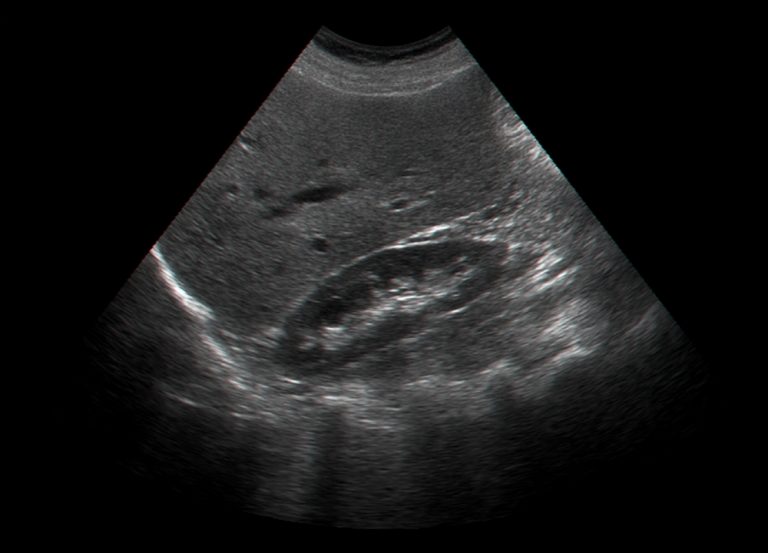
Breast cancer is one of the most common cancers among women, and early detection is essential for effective treatment. Breast ultrasound has become a valuable tool in the diagnosis and assessment of breast cancer, offering non-invasive imaging that provides detailed information without the need for radiation. In London, private ultrasound services have expanded access to this important diagnostic option, giving patients timely evaluations with enhanced convenience. Here’s an in-depth look at what breast ultrasound offers and how it plays a role in diagnosing breast cancer.

What is Breast Ultrasound?
Breast ultrasound is an imaging technique that uses high-frequency sound waves to create visual images of the internal structures of the breast. Unlike mammography, which uses X-rays, ultrasound relies on sound waves to produce images, making it a radiation-free option. During an ultrasound, a technician or radiologist applies a gel to the skin and moves a small transducer over the area to capture images. This procedure allows for real-time assessment of breast tissues, and because it doesn’t use radiation, it is safe for repeated use if needed.
Breast ultrasound is particularly useful for examining dense breast tissue, which can sometimes limit the effectiveness of mammograms. According to the American Cancer Society, women with dense breasts are at a slightly higher risk of developing breast cancer. For these women, ultrasound is an essential supplemental tool as dense tissue can obscure abnormalities in mammograms. Ultrasound offers a clearer view of such tissue, helping identify areas of concern that may require further examination.
How Breast Ultrasound Assists in Breast Cancer Diagnosis
Breast ultrasound excels at providing detailed images that can distinguish between different types of breast abnormalities. It allows radiologists to differentiate between solid masses, such as tumours, and fluid-filled cysts, which are generally benign. This ability to characterize lumps is crucial in breast cancer diagnosis, as it helps in determining which abnormalities are likely to require biopsy or further testing.
Studies published in the American Journal of Roentgenology have shown that breast ultrasound is highly sensitive in identifying small and non-palpable breast lesions, making it particularly useful for early detection. Research also shows that combining ultrasound with mammography significantly increases the sensitivity of breast cancer screening in women with dense breast tissue, helping to catch cancers that might otherwise go undetected in mammograms alone.
While mammography is often the first imaging step in breast cancer screening, ultrasound is valuable when mammogram results are inconclusive. It serves as a second layer of analysis, giving doctors a more detailed view of any suspicious areas. In cases where a mammogram detects a questionable lesion, ultrasound can provide additional clarity on whether it appears malignant, benign, or complex, which may guide the next steps in diagnosis.
When Breast Ultrasound is Recommended
Breast ultrasound is commonly recommended in specific situations where additional clarity is required. It is often used when there is a palpable lump that needs further evaluation. It’s also frequently recommended for women who experience breast pain, unexplained nipple discharge, or other symptoms that could indicate an underlying issue. For younger women under the age of 40, whose breast tissue is generally denser, ultrasound is often preferred over mammography as an initial imaging test.
For women with a known benign condition like fibroadenomas, ultrasound is an effective way to monitor the condition over time, reducing the need for more invasive procedures. A study published in Radiology highlighted that ultrasound follow-up in patients with benign breast disease significantly reduced unnecessary biopsies and invasive procedures, promoting a more conservative approach to managing non-cancerous conditions.
In patients who have undergone treatment for breast cancer, ultrasound can be a valuable tool for monitoring any changes in the breast tissue post-treatment. Regular ultrasound exams can help in detecting any recurrent abnormalities at an early stage, providing peace of mind for those concerned about the possibility of recurrence.

The Process of a Breast Ultrasound Appointment
A breast ultrasound appointment is generally straightforward and non-invasive. The procedure is typically completed in 15 to 30 minutes, and no specific preparation is required. Patients lie on an examination table, and the radiologist or sonographer applies a gel to the breast area. This gel helps transmit sound waves from the ultrasound probe into the breast tissue. The probe is then moved across the breast, and real-time images appear on a monitor.
Radiologists look for specific characteristics in the images to interpret the results. For example, they may assess the shape, edges, and composition of any lumps or cysts they find. Rounded, smooth-edged lumps often suggest benign conditions, while irregular shapes or indistinct edges might indicate malignancy. For patients, understanding the process can alleviate some of the anxiety that may come with an ultrasound appointment, as the procedure is both quick and painless.
Ultrasound is also increasingly popular for guiding biopsies of suspicious lumps. By pinpointing the exact location of a lesion, ultrasound-guided biopsies enable radiologists to obtain tissue samples with high accuracy and minimal discomfort. This targeted approach is especially valuable for lesions that are difficult to locate with other imaging techniques, ensuring that patients receive an accurate diagnosis without invasive surgery.
Advantages of Choosing Private Breast Ultrasound Services in London
In a city like London, private ultrasound services offer numerous benefits for patients seeking breast ultrasound exams. One of the primary advantages is the reduced waiting times, allowing for faster appointments and quicker access to diagnostic results. For individuals who may be experiencing worrying symptoms or are at higher risk, quick access to imaging can provide reassurance or enable prompt treatment.
Private services often provide continuity of care, with patients frequently seeing the same specialists across appointments, leading to a more personalized experience. These facilities often employ highly experienced radiologists with specialized training in breast imaging, ensuring that patients receive the most accurate interpretations of their ultrasound results. Many private ultrasound centers also offer same-day reporting, which can be invaluable for individuals seeking prompt answers.
Furthermore, private clinics in London are equipped with advanced imaging technologies, ensuring high-quality images that facilitate accurate diagnoses. This level of technology is particularly important in breast ultrasound, where precise imaging of small or subtle changes in breast tissue can make a significant difference in the diagnostic process.
Using Breast Ultrasound to Guide Biopsies
Ultrasound’s role extends beyond diagnostics to assist in biopsy procedures. Ultrasound-guided biopsy is a minimally invasive procedure that uses real-time imaging to guide a needle to the exact site of an abnormality, allowing for accurate tissue sampling. Research in the European Journal of Radiology shows that ultrasound-guided biopsies yield high accuracy rates, providing a reliable means of confirming or ruling out breast cancer without the need for surgical intervention.
In London’s private ultrasound clinics, this procedure is typically performed as an outpatient service, enabling patients to go home the same day. During the biopsy, local anesthesia is used to ensure patient comfort, and the ultrasound guidance minimizes the chances of sampling error. With results often available within a few days, patients experience a streamlined diagnostic process, which is particularly beneficial in high-stress situations where a quick diagnosis is essential.
Understanding the Limitations of Breast Ultrasound
While breast ultrasound is a highly effective diagnostic tool, it is important to understand its limitations. Ultrasound may not detect all types of breast cancers, particularly in cases where a lesion is very small or lacks clear boundaries. It is typically used as a supplemental imaging technique rather than a standalone method, especially for women at higher risk.
For patients with dense breasts, combining mammography with ultrasound provides a comprehensive approach to screening. Studies indicate that combining the two increases the detection rate of cancers compared to using either method alone. MRI is sometimes also recommended as an additional tool for high-risk patients. This layered approach to screening helps ensure that any abnormalities are thoroughly investigated, reducing the likelihood of missed diagnoses.
Conclusion
Breast ultrasound is a valuable tool in the diagnosis of breast cancer, especially for women with dense breast tissue or symptoms that require further investigation. With its ability to distinguish between different types of lumps and guide biopsies, ultrasound provides a precise, non-invasive option that complements other imaging techniques. For patients seeking private ultrasound services in London, access to this technology means timely and personalized care, making it easier to navigate the often complex journey of breast health diagnostics.
References:
- American Cancer Society. (2021). Breast Cancer Early Detection and Diagnosis. Retrieved from https://www.cancer.org/cancer/breast-cancer/screening-tests-and-early-detection.html
- American College of Radiology. (2021). Ultrasound - Breast. Retrieved from https://www.acr.org/-/media/ACR/Files/Radiology-Safety/Radiology-Info/Ultrasound_Breast.pdf
- National Cancer Institute. (2021). Breast Cancer Screening. Retrieved from https://www.cancer.gov/types/breast/screening-fact-sheet
- National Health Service. (2021). Breast ultrasound. Retrieved from https://www.nhs.uk/conditions/breast-cancer-screening/breast-ultrasound/
- Radiological Society of North America. (2021). Ultrasound - Breast. Retrieved from https://www.radiologyinfo.org/en/info.cfm?pg=breastus
Content Information
We review all clinical content annually to ensure accuracy. If you notice any outdated information, please contact us at info@iuslondon.co.uk.
About the Author:

Dr. Mohammad Jalaleddin (GMC number: 7729988) is an internationally-trained physician who earned his medical degree from Tishreen University in Syria in 2005. He completed his specialization in diagnostic radiology in 2009, followed by a Master's degree in 2011, which focused on the radiological alterations of bone grafts. Dr. Jalaleddin has worked as a radiologist in various hospitals and clinics, developing significant expertise in ultrasound and MRI scans. He has extensive experience in performing a wide range of ultrasound examinations, including breast, abdominal, pelvic, and vascular studies. In 2017, he moved to the UK and completed the requalification process with the General Medical Council (GMC). Since March 2021, he has been a part of the Imperial College Healthcare NHS Trust. Dr. Jalaleddin is a member of the General Medical Council (GMC) and the British Medical Ultrasound Society (BMUS).


