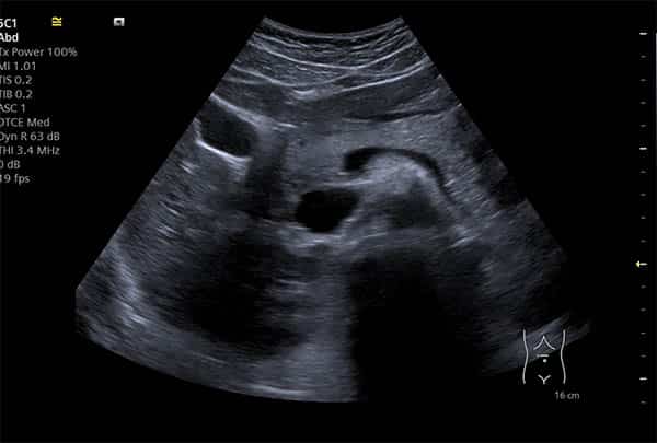
Ultrasound imaging, or sonography, is a non-invasive medical diagnostic procedure that uses ultrasonic sound waves to create images of the body. These images are used by physicians to diagnose and treat various medical conditions. A patient may have an ultrasound for a variety of reasons, such as pregnancy, organ or soft tissue evaluation and assessment of blood vessels. This guide provides an overview of ultrasound exams, including the types of examinations available, what patients can expect during the exam and how to interpret ultrasound results.
Ultrasound imaging typically does not require any sort of sedation or radiation exposure. It’s a safe procedure which can produce detailed pictures of organs, lesions and other structures inside your body through harmless sound waves bouncing off them. Ultrasound can even provide real-time images in motion such as beating heart or functioning organs like kidneys or gallbladder. During an ultrasound examination, you may be asked to move into different positions so that different parts of your body can be analysed at different angles to obtain more detailed images.
What is an Ultrasound?
An ultrasound is a medical imaging technology which uses sound waves to create a detailed image of the inside of your body. This image can help your doctor diagnose any abnormalities or identify any structural changes on the organs, tissues and veins. Ultrasounds are a non-invasive, painless procedure that can provide your doctor with important information about your health.
Let's explore further to understand how this imaging technology works and how it can be used for diagnosis:
Types of Ultrasounds
Ultrasound is a non-invasive medical imaging technique used to observe the internal structures of the body. It works by sending sound waves which are reflected (echoed) off internal structures back to a device which then produces an image of whatever is being examined. Ultrasound exams can be used to check organs and soft tissue, as well as bones in certain cases. The type of sound wave used, its frequency and intensity determines what type of information will be detected.
There are several different types of ultrasounds that may be used in clinical practice:
- B-mode ultrasound: This type of imaging uses only one frequency, allowing for 2D images to be created that usually depict only grey tones. B-mode ultrasound is commonly used for fetal imaging.
- Doppler ultrasound: Doppler ultrasound uses higher frequencies and has the ability to detect movement and directionality in blood cells or other moving objects, such as an unborn baby’s heart rate or blood flow through an organ. Doppler studies have a characteristic “shiny” look compared to standard B-mode ultrasound images because they contain spectral energy information from movement within the tissue being imaged. This type of imaging is particularly useful for assessing foetal health and monitoring in pregnancy, as well as studying blood flow within vessels for atherosclerosis and arterial insufficiency risk assessment.
- 3D/4D Ultrasound: These types of ultrasounds use multiple frequencies to produce 3D images which can show static structures or developing foetuses over time (ultrasound 4D). These images yield much more detailed results than standard 2D B-mode videos typically obtained during routine scans; they are utilized primarily when complex foetal anatomy needs further evaluation or when expecting parents want a life like image earlier on in the pregnancy than traditional 3D technology can capture.
Preparation for the Test
Prior to your ultrasound, it is important that you receive detailed information about what the exam will entail. You may be asked to wear a gown or special clothing for the procedure, and you should be aware of any modifications or contraindications for your situation. Your doctor may advise that you fast or limit food and fluids prior to the ultrasound examination. Make sure to discuss any recent medical tests, medications (over-the-counter and prescription) taken in the last 24 hours, or changes in your health over the past few weeks with your radiologist before beginning an ultrasound.
In some cases, a patient may need to drink water or other fluids prior to an abdominal scan. In other cases a full bladder is necessary for certain examination types such as obstetric and gynaecologic ultrasounds. Additionally, if you have had a contrast material used in another imaging study recently, the radiologist should be made aware of this before the ultrasound begins. Women should also tell their technician if they are pregnant as some precautions might need to be taken during their examination depending on which area of their body is being imaged.
For most ultrasounds there is no additional preparation necessary; however it’s important that you follow your doctor’s instructions leading up to your exam so that an accurate diagnosis can be achieved.
What to Expect During an Ultrasound
An Ultrasound is a non-invasive procedure that uses sound waves to create images of the body’s internal organs. It is a painless procedure that can provide your doctor with important information about your health.
During an Ultrasound, your doctor or technician will use a transducer to create images of the organs or structures they are looking to examine. The transducer emits sound waves and then listens for the echoes that bounce off your body.
Ultrasound Procedure
During an ultrasound procedure, a technologist will use a transducer or probe to make images of the inside of your body. This probe is moved across your skin, which may appear as if it’s being scanned with a light. Ultrasound waves are then sent from the transducer into the area being examined. The sound waves interact with soft tissues, organs and blood vessels in your body. The reflections of these sound waves are detected by the transducer and relayed by computer to form images that can be seen on a monitor.
In some cases, a gel will be applied to your skin prior to the ultrasound being performed. This gel allows for better contact between the skin and transducer so that sharper images can be produced. Once completes, the gel will simply need to be wiped off. Depending on what area is being examined and its size, some wider probes may also be used during an ultrasound procedure so that larger structures can be visualized more clearly.
Ultrasound Images
During an ultrasound procedure, sound waves will be passed through the area of the body being examined. These waves are reflected off of various objects and structures, such as tissue and organs. In order to create images on the monitor of what is inside the patient’s body, these echoes are then processed and reconstructed into live video images. These images allow for a real-time understanding of the anatomy that may be too deep for other imaging technology to reach in such detail.
The examination begins with a general scan to obtain an overall view from which particular areas can then be studied more closely if required. The specific type and orientation of transducer used depend on what area it will probe – e.g., abdominal and chest exams use a convex transducer – and where within that area it will radiate ultrasonic impulses (a linear transducer is usually used for abdominal scanning as this allows for higher resolution). In real-time mode, images are constantly refreshed as different views are achieved by changing the angle or depth of the beam directed by your sonographer (ultrasound technician).
It is also possible to take still pictures using "digital capture". Using this method, an image can be frozen on screen in both 2D format (regular ultrasound view) or 3D format using special 3D probes that further enhance details seen onscreen. Once captured onto disk, these images can then be stored, printed or emailed to specialists at other locations if needed.
Understanding Ultrasound Results
Ultrasound scans are important tools used by medical professionals to diagnose medical conditions and assess a patient's health. An ultrasound scan records sound waves that reflect off of internal organs and structures, providing a picture of the area being examined.
Thanks to modern technology, patients can easily understand the results of their ultrasound scan. This guide provides an overview of how to properly interpret and understand the results of an ultrasound scan:
Normal Ultrasound Results
A normal ultrasound result indicates that no abnormalities were found in the imaging. A normal result does not rule out all possible medical conditions, as some diseases may not show up clearly on an ultrasound. However, in general, these results point to a higher chance of good health.
When interpreting ultrasound results, it is important to remember that what looks normal for one person may not look normal for another; depending on the scan and the patient's age and gender, different structures may be visible for evaluation. Additionally, a few factors can affect how well the ultrasound displays certain structures such as gestation age or body composition level.
Normal ultrasound readings create a reference point for future scans and help the practitioner monitor any changes that might occur within the patient’s body over time. Examples of normal readings include:
- Expected size and placement of organs
- No evidence of fluid build-up or swelling
- No sustained increases in temperature
- Normal tissue density throughout the imaging area
- Normal shapes and sizes of blood vessels
- Normal patterns of blood flow
Abnormal Ultrasound Results
When an ultrasound diagnosis indicates that something is wrong, it can be a stressful time for the patient and their family. Patients should understand three important terms when discussing ultrasound results: “abnormal”, “suspect”, and “indeterminate”.
- Abnormal result means there is a problem that requires medical attention and usually further testing. It could indicate a major abnormality such as an aneurysm of the abdominal aorta or a congenital problem such as hypoplastic left heart syndrome in an unborn baby.
- Suspect results are essentially inconclusive and may require additional testing to make a diagnosis. The suspect term is often used in foetal ultrasounds to describe findings that require further examination before proceeding with treatment options such as antibiotics for prenatal infections or karyotyping for chromosome abnormalities.
- Indeterminate results mean the ultrasound failed to demonstrate an abnormality but cannot rule it out either. This often occurs when the evidence of disease was insufficiently visualized, either due to patient anatomy or imaging technique errors in obtaining quality images through organs or body cavities which do not allow direct visualization. Indeterminate results may require additional imaging on another type of equipment or closer follow up over time to determine if there has been any resolution of the issue at hand.
Follow Up Tests
After receiving your ultrasound results, you may have unresolved questions or require further investigative tests for a proper diagnosis. Follow up tests can be recommended for a variety of reasons. Depending on your ultrasound results, your doctor may order additional imaging tests such as x-rays, MRIs, CT scans or ultrasounds with Doppler to provide more detailed pictures of the body parts in question. Imaging tests can also be repeated over time to compare the changes in affected organs and tissues.
Your doctor may also recommend laboratory tests such as blood work to asses internal organ function or determine the presence of an infection or abnormal levels of chemicals and minerals in the body to ensure better understanding of the initial results. Endoscopic examinations are diagnostic procedures done internally that can provide a more detailed view inside the body without invasive surgery. Additional follow-up appointments may include medical consultation with a specialist who specializes in conditions related to your specific diagnosis and ongoing care plans related to complex matters that could not be answered by initial imaging tests.
Conclusion
As you can see, there is a lot to consider when discussing your ultrasound results. It’s important to maintain open communication with your healthcare provider and ask questions about any of the findings before making a final diagnosis or treatment decision.
Your provider will work with you to review the ultrasound images and provide an interpretation that is specific to your situation. With proper understanding, you can rest assured knowing that your medical team will utilize the best available diagnostic technique when guiding you through a course of care.


