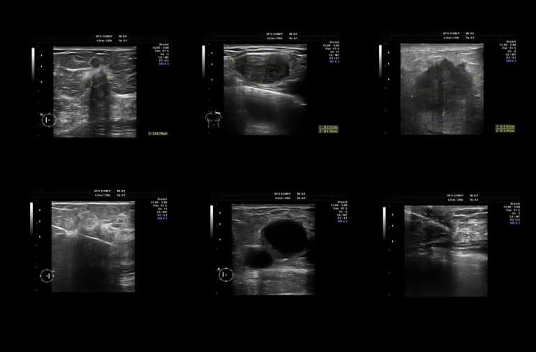
What is a thyroid ultrasound?
Ultrasound imaging, also called ultrasound scanning or sonography, involves the use of a small transducer (probe) and ultrasound gel to expose the body to high-frequency sound waves. It is safe and painless and produces pictures of the inside of the body using sound waves. Ultrasound imaging is a non-invasive medical test that helps physicians diagnose and treat medical conditions.
The thyroid gland is located in front of the neck just above the collar bones and is shaped like a butterfly. It is one of nine endocrine glands that make and send hormones into the bloodstream.
The thyroid gland makes the thyroid hormone, which helps to regulate a variety of body functions, including how fast the heart beats. Commonly, patchy areas or nodules develop in the thyroid that may or may not be felt on the skin surface. These are called palpable nodules.
Ultrasound is very sensitive and shows nodules that cannot be felt. The vast majority of these are benign regions of thyroid tissue that pose no health risk, but a few are true tumours of the thyroid and may require further diagnosis or treatment. You can read more about the symptoms of thyroid cancer.
What are some common uses of the procedure?
An ultrasound of the thyroid is typically used to:
· to determine if a lump in the neck is arising from the thyroid or an adjacent structure
· to analyse the appearance of thyroid nodules and determine if they are the more common benign nodule or if the nodule has features that require a biopsy
· to look for additional nodules in patients with one or more nodules felt on physical exam
· to see if a thyroid nodule has substantially grown over time
Because ultrasound provides real-time images, images that are renewed continuously, it also can be used to guide procedures such as needle biopsies. Ultrasound may also be used to guide the insertion of a catheter or other drainage device.
How should I prepare?
You should wear comfortable, loose-fitting clothing for your ultrasound exam. You may need to remove all clothing and jewellery in the area to be examined, and you may be asked to wear a gown during the procedure. No other preparation is required.
How is the procedure performed?
For most ultrasound exams, the patient is positioned lying face-up on an examination table that can be tilted or moved. A pillow may be placed behind the shoulders to extend the area to be scanned.
A clear water-based gel is applied to the area of the body being studied to help the transducer make secure contact with the body and eliminate air pockets between the transducer and the skin that can block the sound waves from passing into your body. The sonographer (ultrasound technologist) or radiologist then presses the transducer firmly against the skin in various locations.
When the examination is complete, the patient may be asked to dress and wait while the ultrasound images are reviewed.
The ultrasound examination is usually completed within 30 minutes and is painless, fast and easily tolerated by most patients.
If scanning is performed over an area of tenderness, you may feel pressure or minor pain from the transducer.
You may need to extend your neck to help the sonographer to examine your thyroid with an ultrasound. If you suffer from neck pain, inform the sonographer so that they can help situate you in a comfortable position for the exam.
Once the imaging is complete, you are able to resume your normal activities immediately.
What are the benefits vs. risks?
Benefits
· Most ultrasound scanning is noninvasive (no needles or injections).
· Occasionally, an ultrasound exam may be temporarily uncomfortable, but it is almost never painful.
· Ultrasound is widely available, easy-to-use and less expensive than other imaging methods.
· Ultrasound imaging is extremely safe and does not use any ionizing radiation.
· Ultrasound scanning gives a clear picture of soft tissues that do not show up well on x-ray images.
· Ultrasound provides real-time imaging, making it a good tool for guiding minimally invasive procedures such as needle biopsies and needle aspiration.
Risks
· For standard diagnostic ultrasound, there are no known harmful effects on humans.
If you are suspected thyroid issues you can book a private ultrasound in our ultrasound clinic in London instead of waiting sometimes more than 6 weeks for an NHS scan.


