Ultrasound scans during your pregnancy are an invaluable way to learn more about your unborn child and can also help doctors detect any problems with the uterus, ovaries, or cervix.
Ultrasounds can be obtained on the NHS or privately for a fee. However, there are some things to consider before choosing to opt for private ultrasounds.
What is an Ultrasound Scan?
Ultrasound scans are non-invasive tests that use sound waves to create a real-time image of inside your body. Ultrasound can help diagnose various conditions and is commonly employed during pregnancy to monitor the health of an unborn baby.
The technology works by sending high frequency sound waves directly at the area of your body that needs examination. As these sound waves bounce off the organs and bounce back to the probe, which detects their echoes and builds a picture of that part of your body.
Sonographers, a type of healthcare professional, use ultrasound to check your heart, lungs, bladder and other body areas. They apply gel onto your skin before placing a probe on top. The gel helps bounce sound waves off tissue back towards their source at the probe.
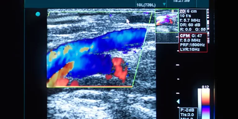
They will then maneuver the probe around your body to get a clear image of what lies within. They may also use Doppler ultrasound technology, which allows them to assess blood flow through your arteries and veins.
Doppler ultrasounds can detect a range of conditions, such as blockages in blood flow or narrowed vessels in your heart or lungs that could indicate an infection or cardiovascular problem such as an artery clogged with fat, heart valve issues or plaque buildup. They may even determine the source of symptoms like lumps if they’re cancerous tumours or fluid-filled cysts.
Transvaginal ultrasound pictures take pictures of your ovaries, womb and surrounding structures while you lie flat on an examination table. Although it won’t hurt you, please inform the sonographer if it causes any discomfort for you.
Before your test, it’s wise to empty your bladder. Additionally, drink plenty of water as you may be instructed by the doctor or sonographer to do so.
Ultrasound scanning is a quick, painless procedure that doesn’t involve radiation and usually feels comfortable – although some people may feel some pressure from the probe being placed on their skin. You’ll usually be able to go home or back to work immediately after your ultrasound scan, with results available within a few days.
How Does an Ultrasound Scan Work?
Ultrasound is a medical imaging technique that utilizes high-frequency sound waves to create an image of part of your body. It’s often employed to monitor pregnant women, diagnose diseases and guide surgeons during certain procedures.
Ultrasound scans involve the use of a small transducer (probe) that transmits sound waves into the body and records them when they bounce back or echo off different surfaces. A computer then interprets these echoes to produce an immersive two-dimensional image onscreen; higher the frequency of these sound waves, the clearer and clearer it appears.
Most types of diagnostic ultrasound involve placing the probe on your skin. Certain exams, like transvaginal scans, require that the probe be placed inside your body.
The ultrasound technician will apply a thin layer of gel onto the area being scanned. This gel helps transmit sound waves and prevents air pockets from forming between the probe and skin. They then move the probe over this gel in order to capture an image of what’s being scanned.
Once the technician has taken enough pictures, they will wipe away any gel and your doctor can review the images with you. Generally, results of an ultrasound exam can be seen within a few hours after it has been concluded.
Ultrasound images taken during pregnancy can be useful to determine due dates and rule out ectopic pregnancies and other potential health problems. They also allow us to monitor fetal growth and check on breech positioning, among other things.
Ultrasound can also be used for elastography, which measures tissue stiffness and helps doctors distinguish tumors from healthy tissue. This information can be displayed as either color-coded maps of relative stiffness or black-and-white maps that show high contrast images of tumors compared with anatomical images.
Ultrasound can also be used medically to visualize internal organs and examine blood flow in certain regions of the body, like arteries and veins. In certain instances, ultrasound can even be used to diagnose kidney stones or calcification of blood vessels. Furthermore, it has the capability to detect cysts and other growths within a woman’s womb or pelvic area as well as elsewhere on her body.
What Are the Benefits of a Private Ultrasound Scan?
Ultrasound is a test that utilizes high-frequency sound waves to produce images of your body’s internal organs and soft tissues. Unlike X-rays, ultrasound uses no radiation and is safe for people of all ages – including pregnant women!
This test is painless and doesn’t involve needles or cuts in your skin. Instead, a transducer sends pulses of sound waves through gel onto your skin, which are converted into electrical signals displayed on a computer screen. As these waves travel into body tissue and create echoes, the probe converts them into electrical signals for analysis.
Another advantage of an ultrasound is that it does not require contrast agents, which some imaging tests require to highlight certain issues. This makes it safer for people with allergies or sensitivity to contrast dyes or who have had prior reactions with them.
Additionally, it offers accurate results without taking up unnecessary time or space in your home. Many private providers provide affordable and low-cost examinations so you can get the information needed to make informed decisions about your health care.
Ultrasound can provide you with peace of mind during your pregnancy. The procedure can also detect birth defects, breech positioning and placental issues. By having an ultrasound early on in your pregnancy, you can take preventative measures to help avoid future issues and ensure that your baby is growing normally and securely.
An ultrasound can also reveal if you’re expecting twins, adding an extra layer of excitement to your pregnancy. It also helps determine the gender of your baby during the first trimester, so you’ll be ready to share the news with all of your loved ones!
If you have any doubts about whether an ultrasound is suitable for you, consult with your doctor or midwife. They can explain the advantages of the test and help determine if it meets your requirements.
It’s essential to respect your privacy during an ultrasound scan. Selecting a provider who will treat you with respect and offer the best experience possible is key. A private ultrasound clinic typically has cutting-edge equipment in a professional setting, ensuring your scan runs smoothly without feeling rushed or uncomfortable during it. This way, you can ensure the most comfortable experience possible during your scan.
What Are the Drawbacks of a Private Ultrasound Scan?
Private ultrasound clinics typically boast state-of-the-art facilities that are clean and tastefully decorated. The doctors and technicians are highly experienced to perform procedures quickly. Furthermore, they are familiar with their machines so they can identify any complications in images quickly.
Ultrasound imaging is a safer alternative than x-rays or CT scans, as it doesn’t use radiation. It has become one of the most commonly used and popular ways for pregnancy care providers to screen women for conditions that could harm their babies’ health.
Ultrasounds can also help your doctor detect certain birth defects and medical conditions, such as cleft palate, that might be caused by problems with your baby’s brain, heart or bones (congenital disorders). This is an essential part of prenatal care because it gives you insight into your baby’s condition before they arrive at the hospital.
Your pregnancy care provider uses abdominal or transvaginal ultrasound to capture images of your baby’s face and body. These usually 2D images can also be enhanced with 3D or 4D ultrasounds for better views of the head, arms, legs and organs.
After 12 weeks of gestation, your provider can perform prenatal ultrasound scans to capture images of your baby’s organs and other parts of the abdomen using a transducer device. They apply slight pressure for the best view possible during this procedure.
Ultrasound scans take around 20 minutes and should not be painful. However, you should drink a lot of water and refrain from drinking or urinating two hours prior to your ultrasound appointment.
Your doctor may need to do a follow-up ultrasound exam to determine whether the issue identified in your first ultrasound has resolved or not. This examination also offers them additional views or utilizes special imaging techniques for more comprehensive assessment of the condition.
When scheduling an ultrasound, the amount of time that should pass between your first and second tests depends on both the type of scan you receive and any medical complications. Always consult your obstetrician or general practitioner for further advice about whether or not an ultrasound is necessary for you.


