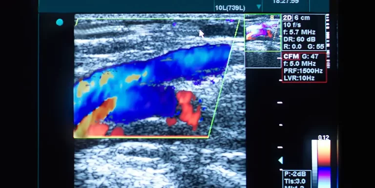
Abdominal health is central to overall well-being, as it affects core body functions and many essential organs. When dealing with unexplained discomfort, digestive problems, or other abdominal concerns, an ultrasound scan often becomes the first step in diagnosis. Abdominal ultrasound is a non-invasive imaging technique that uses high-frequency sound waves to create detailed images of organs and structures within the abdomen. It offers a safe, radiation-free method for diagnosing a wide range of health conditions.
For those considering a private ultrasound, this guide outlines the entire process, what to expect, and how it can be a critical step in diagnosing and managing abdominal health issues.

What Is an Abdominal Ultrasound?
An abdominal ultrasound, also known as a sonogram, utilizes sound waves to produce real-time images of internal structures. Unlike X-rays or CT scans, ultrasound does not involve radiation, making it a safe option for regular imaging. The procedure is relatively simple and is typically performed by a trained sonographer, who uses a hand-held device called a transducer. This device emits sound waves that bounce off tissues and organs, creating images on a screen.
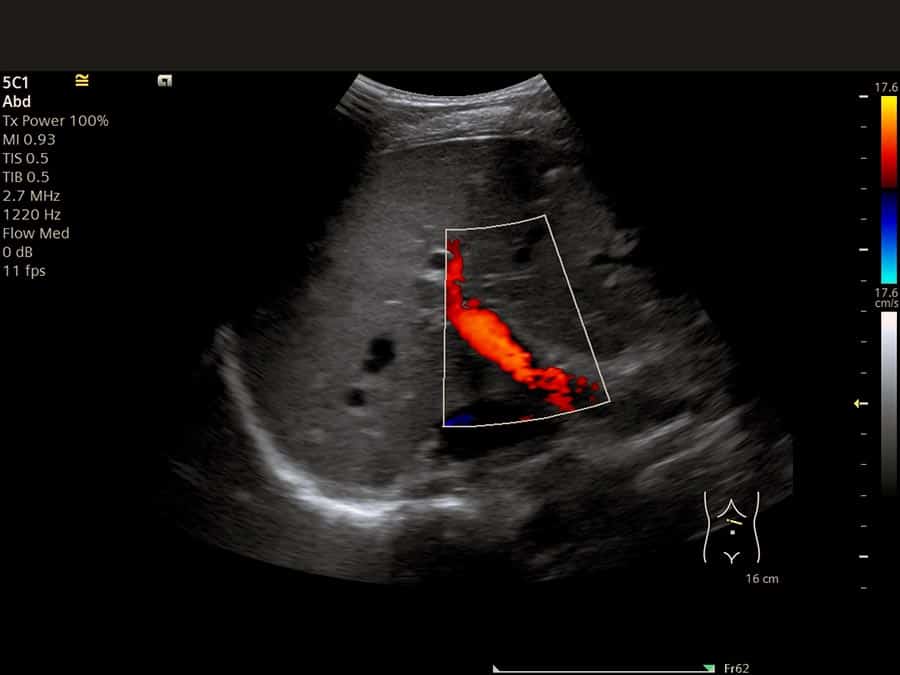
Abdominal ultrasounds are commonly used to examine major organs within the abdominal cavity, including the liver, kidneys, pancreas, gallbladder, spleen, and abdominal blood vessels. They can detect abnormalities such as cysts, tumors, blockages, and infections, as well as signs of inflammation or organ enlargement.
Common Health Conditions Diagnosed with Abdominal Ultrasound
Abdominal ultrasounds play a vital role in diagnosing a broad range of health conditions that affect various organs. Here are some of the most common issues that can be detected through this imaging method:
-
Digestive System Disorders
The liver, gallbladder, and pancreas are essential organs in the digestive system, each with its own function in processing and distributing nutrients. Liver disease, such as fatty liver or cirrhosis, can be effectively detected through ultrasound, as can gallstones, which form in the gallbladder and can block bile flow, causing pain. A study published in Hepatology International emphasized that ultrasound is often the first-choice imaging technique for diagnosing fatty liver disease, given its accuracy and non-invasive nature. Additionally, pancreatitis or pancreatic tumors can also be identified on an ultrasound scan. -
Urinary and Kidney Conditions
The kidneys and bladder are also visible on an abdominal ultrasound, allowing for the detection of urinary system disorders like kidney stones or infections. According to research in the Journal of Urology, ultrasound is often the preferred method for diagnosing kidney stones, particularly in cases where non-invasive imaging is beneficial. Other kidney-related issues, such as cysts or structural abnormalities, can also be assessed with ultrasound. -
Vascular Health Issues
Ultrasound is invaluable in identifying vascular abnormalities within the abdomen, such as abdominal aortic aneurysms (AAA). AAA, a potentially life-threatening condition where the main abdominal artery swells or weakens, can be identified early through ultrasound. A study in Vascular Surgery reports that ultrasound screenings are instrumental in reducing AAA mortality by enabling timely diagnosis and intervention. -
General Abdominal Pain and Unexplained Symptoms
Abdominal pain is a common reason for seeking an ultrasound. Ultrasound helps pinpoint the underlying cause, whether it’s a benign cyst, an inflamed appendix, or even less common issues like an abdominal hernia. Doctors use ultrasound to visualize organs in real-time, allowing them to quickly assess the cause of pain and recommend a course of treatment.
Preparing for Your Abdominal Ultrasound
Proper preparation for an abdominal ultrasound can ensure clearer images and a smoother experience. Most ultrasounds require simple steps, such as fasting for several hours beforehand. Fasting minimizes gas in the intestines, which could obstruct the ultrasound waves and reduce the clarity of the images. If a patient needs to undergo a bladder scan, drinking water beforehand may also be necessary to fill the bladder for better visualization.
Wear comfortable clothing that can be easily adjusted or removed to allow access to the abdomen. Additionally, bringing previous medical records, especially any prior imaging reports, can provide context to the sonographer or radiologist performing the scan.
What Happens During an Abdominal Ultrasound?
An abdominal ultrasound procedure is straightforward and typically lasts around 20-30 minutes. The patient will lie on an examination table, and the sonographer will apply a clear, water-based gel to the abdomen. This gel aids the transmission of sound waves, helping the transducer capture clearer images. The sonographer then moves the transducer across different areas of the abdomen, capturing images of each organ in detail.
Patients might feel mild pressure from the transducer, especially over areas that are tender or sensitive, but the procedure is generally painless. Since the images are captured in real-time, the sonographer may ask the patient to hold their breath briefly to get clearer images, particularly around the liver and spleen, which are sensitive to the movement caused by breathing.
After the Ultrasound: Interpreting the Results
Once the ultrasound is complete, the sonographer or radiologist will analyze the images and generate a report, which is typically sent to the patient’s referring doctor within a few days. Results may include information on the size, shape, and texture of organs, any detected abnormalities, and signs of inflammation or disease.
For example, the liver might appear enlarged if there is a sign of liver disease, or gallstones may be visible within the gallbladder. If any abnormal growths, cysts, or unusual fluid collections are identified, the report will indicate their characteristics, aiding the physician in determining whether further tests are necessary.
Benefits of Choosing a Private Ultrasound Service
Private ultrasound services are becoming increasingly popular due to the faster access to diagnostic imaging they provide. Unlike public health systems, where patients may experience long wait times, private clinics often allow patients to schedule an appointment promptly. This can be particularly beneficial for those experiencing ongoing discomfort or symptoms that require swift diagnosis.
Private clinics also tend to offer a more personalized experience, often giving patients more time to discuss their symptoms and health history with their radiologist. These clinics may utilize advanced ultrasound technology, which can provide high-resolution images and more detailed diagnostic information.
Research highlighted in The Journal of Diagnostic Imaging shows that patients report higher satisfaction with private ultrasound services due to these benefits, including the opportunity for a more comprehensive explanation of findings and recommendations for next steps in their healthcare journey.
Frequently Asked Questions (FAQs)
Is an abdominal ultrasound painful?
No, abdominal ultrasounds are non-invasive and generally painless. Some patients may feel mild pressure when the transducer is moved over sensitive areas, but discomfort is minimal and temporary.
Do I need a referral for a private ultrasound?
Many private ultrasound clinics accept self-referrals, meaning patients can directly book an appointment without a doctor’s referral. However, some clinics may still require a referral for certain types of scans, so it’s best to check with the specific clinic in advance.
How accurate are abdominal ultrasound results?
Ultrasound is highly effective for visualizing soft tissues and fluid-filled structures. Studies, including those published in Radiology, confirm ultrasound’s reliability for diagnosing conditions like gallstones, liver disease, and kidney issues. However, for more complex conditions or where more detail is required, additional imaging techniques such as CT or MRI might be recommended.
Will insurance cover the cost of a private ultrasound?
Coverage varies depending on the insurance provider and the patient’s specific plan. Private ultrasound services are generally considered out-of-pocket expenses unless specified by the insurance company as necessary for diagnosis. Contacting the insurance provider for clarification on coverage is recommended.

