Heart health is paramount, and understanding the tools used to monitor it can be immensely helpful. Echocardiography, or simply an echo, is a non-invasive imaging technique that uses ultrasound waves to create detailed images of the heart. This powerful diagnostic tool helps cardiologists assess the structure and function of the heart, ensuring accurate diagnoses and effective treatment plans.
Echocardiography offers several types, each suited for different diagnostic needs. Transthoracic echocardiography (TTE) is the most common, involving a transducer placed on the chest to send ultrasound waves through the heart. Transesophageal echocardiography (TEE) provides clearer images of the heart’s structures by inserting a specialized probe down the esophagus. Stress echocardiography evaluates how the heart performs under physical stress, while Doppler echocardiography measures the speed and direction of blood flow within the heart.
The process of an echocardiogram is straightforward and typically painless. Patients lie on an examination table while a technician applies a gel to the chest to ensure good contact between the skin and the transducer. The transducer sends ultrasound waves through the chest, which bounce off the heart and create echoes that are translated into detailed images on a monitor. These images help cardiologists identify issues like heart valve disease, cardiomyopathy, and congenital heart defects.
Echocardiography is often recommended when symptoms such as shortness of breath, chest pain, or irregular heartbeats arise. It is a critical tool in the diagnosis and management of many heart conditions. By understanding the heart’s anatomy and functionality through echocardiography, healthcare providers can make informed decisions about patient care.
International Ultrasound Services in Notting Hill, London, offers state-of-the-art echocardiography services, providing patients with accurate diagnoses and comprehensive care. The clinic’s experienced professionals ensure that each patient receives the highest level of service and attention, contributing to better heart health outcomes.
What is Echocardiography?
Echocardiography is a non-invasive imaging technique used to visualize the heart’s structure and function. By utilizing high-frequency sound waves, or ultrasound, echocardiography creates detailed images of the heart, allowing doctors to see how the heart and its valves are functioning in real-time. This diagnostic tool is essential in identifying various heart conditions, from valve disorders to cardiomyopathies.
Different types of echocardiography cater to specific diagnostic needs. Transthoracic echocardiography (TTE) is the most common form, where a transducer is placed on the chest to capture heart images. Transesophageal echocardiography (TEE) involves inserting a specialized probe into the esophagus to obtain clearer images, especially in patients with complex heart conditions. Stress echocardiography combines ultrasound imaging with exercise or medication to evaluate how the heart performs under stress. Doppler echocardiography measures the speed and direction of blood flow, which is crucial in assessing heart valve functions and detecting abnormalities like regurgitation or stenosis.
How Does Echocardiography Work?
Echocardiography works by using ultrasound waves to produce images of the heart. During the procedure, a technician places a transducer on the patient’s chest, which emits high-frequency sound waves that travel through the body and bounce off the heart structures. These sound waves are then captured and translated into live images by the ultrasound machine.
The transducer plays a key role, both sending and receiving the sound waves. The images created can show various heart structures, such as the chambers, valves, walls, and blood vessels. This real-time imaging is vital for evaluating the heart’s pumping strength and detecting abnormalities like fluid around the heart or structural defects.
In transesophageal echocardiography, the process is similar but involves a transducer attached to a thin tube passed down the esophagus. This approach provides higher resolution images, as the esophagus is closer to the heart, offering a clearer view without the interference of the lungs and chest wall.
Doppler echocardiography adds another layer of diagnostic capability by measuring the movement of blood through the heart and vessels. By analyzing changes in the frequency of the ultrasound waves as they reflect off moving blood cells, doctors can assess the speed and direction of blood flow. This information is critical for diagnosing conditions like valve diseases and congenital heart defects.
Studies have shown the effectiveness of echocardiography in diagnosing and managing heart disease. According to research published in the Journal of the American College of Cardiology, echocardiography is highly accurate in assessing cardiac function and detecting heart abnormalities, making it a cornerstone in cardiovascular diagnostics.
When is Echocardiography Recommended?
Echocardiography is a valuable tool for diagnosing and managing heart conditions. It’s commonly recommended when patients exhibit symptoms that might indicate heart issues. For instance, if you’re experiencing unexplained chest pain, shortness of breath, or irregular heartbeats, your doctor might suggest an echocardiogram to investigate further.
Heart murmurs, which are unusual sounds heard during a heartbeat, often lead doctors to order an echocardiogram to determine their cause and significance. In some cases, individuals with a family history of heart disease undergo echocardiography as a precautionary measure.
Echocardiography is also essential for monitoring existing heart conditions. For example, patients with known heart valve disease, heart failure, or congenital heart defects benefit from regular echocardiograms to track their heart’s function and structure over time.
After certain cardiac procedures or surgeries, echocardiography helps doctors evaluate the success of the intervention and ensure there are no complications. It’s also used to assess the effectiveness of treatments for heart conditions, such as medications or lifestyle changes.
The versatility of echocardiography makes it a cornerstone in cardiology, providing critical insights that guide both diagnosis and treatment plans.
Preparing for an Echocardiogram
Preparing for an echocardiogram is straightforward, but there are a few things you can do to ensure the process goes smoothly. Generally, you can eat and drink as usual before a standard transthoracic echocardiogram. However, if you’re scheduled for a transesophageal echocardiogram, fasting for several hours beforehand might be necessary.
Wear comfortable clothing to your appointment. You’ll typically be asked to change into a hospital gown to allow easy access to your chest. The procedure is non-invasive and painless, but understanding what to expect can help ease any anxiety.
During the echocardiogram, you’ll lie on an examination table. The technician will apply a special gel to your chest to enhance the transmission of ultrasound waves. A transducer, which looks like a small microphone, will be moved across your chest to capture images of your heart.
For stress echocardiography, which assesses how your heart functions under physical stress, you’ll either exercise on a treadmill or receive medication to simulate the effects of exercise. It’s best to wear comfortable shoes and clothing suitable for exercise if you’re doing the treadmill test.
Transesophageal echocardiography involves inserting a specialized probe into the esophagus to get detailed images of the heart. This type of echocardiogram provides clearer pictures of certain heart structures. For this procedure, you’ll be given a sedative to help you relax, and your throat will be numbed to reduce discomfort.
After the echocardiogram, you can typically resume your normal activities immediately unless you’ve had a sedative. In that case, arrange for someone to drive you home and take it easy for the rest of the day.
Understanding Echocardiography Results
Echocardiography provides detailed images of the heart, allowing doctors to assess its structure and function. When reviewing these results, several key areas are examined:
Heart Chambers and Walls
The size and thickness of the heart chambers are measured to check for conditions like hypertrophy or dilation. Abnormalities in the heart walls, such as thickening or thinning, can indicate diseases like cardiomyopathy.
Heart Valves
Echocardiography evaluates how well the heart valves open and close. This helps identify valve diseases, such as stenosis (narrowing) or regurgitation (leakage). Clear images can show if blood flow is disrupted, which may require further intervention.
Ejection Fraction and Cardiac Output
Ejection fraction measures how much blood the left ventricle pumps out with each contraction. A normal ejection fraction ranges from 55% to 70%. Lower values may suggest heart failure or cardiomyopathy. Cardiac output, the total volume of blood pumped by the heart per minute, is also calculated to assess overall heart function.
Blood Flow and Pressure
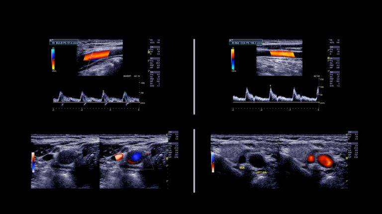
Doppler echocardiography, a specialized type, assesses blood flow through the heart and vessels. It measures the speed and direction of blood flow, helping to detect abnormalities such as turbulence or blockages. Additionally, it provides information on the pressures within the heart chambers, which can indicate conditions like pulmonary hypertension.
Pericardium
The pericardium, the sac surrounding the heart, is also examined. Echocardiography can detect pericardial effusion, an accumulation of fluid around the heart, which might indicate inflammation or infection.
Understanding these aspects helps doctors diagnose a wide range of heart conditions accurately. The detailed information from an echocardiogram is crucial in guiding appropriate treatment plans.
Benefits of Echocardiography
Echocardiography offers several significant benefits, making it a preferred diagnostic tool for heart evaluation:
Non-Invasive and Painless
Unlike some other diagnostic procedures, echocardiography is non-invasive. It uses sound waves to create images of the heart, avoiding the need for incisions or injections. This makes it a comfortable and low-risk option for patients.
Accurate and Detailed Imaging
The technology provides highly accurate and detailed images of the heart’s structure and function. This precision helps in diagnosing a variety of conditions, from valve diseases to cardiomyopathies, ensuring that patients receive accurate and timely treatment.
Real-Time Assessment
Echocardiography allows for real-time visualization of the heart. This capability is especially useful during stress tests or surgeries, as it provides immediate feedback on how the heart is performing under different conditions.
Versatile Applications
It can be used in various settings, including emergency rooms, outpatient clinics, and during surgeries. Its versatility means it can be applied to a wide range of patients, from newborns with congenital heart defects to adults with acquired heart diseases.
Guides Treatment Decisions
The detailed information obtained from an echocardiogram is invaluable in guiding treatment decisions. Whether it’s determining the need for surgery, evaluating the effectiveness of medications, or planning lifestyle changes, echocardiography provides a solid foundation for managing heart health.
Low Risk and Cost-Effective
Compared to other imaging modalities like MRI or CT scans, echocardiography is cost-effective. Its non-invasive nature also reduces the risk of complications, making it a safer choice for many patients.
Risks and Limitations
While echocardiography is a highly valuable tool, it does have some risks and limitations:
Potential Discomfort
Patients might experience slight discomfort from the transducer pressing against the chest, especially if they need to lie still for extended periods. In transesophageal echocardiography, mild throat discomfort or a gag reflex might occur due to the insertion of the probe.
Limited Views in Certain Patients
In some cases, obtaining clear images can be challenging. Factors such as obesity, chronic lung disease, or having a thick chest wall can limit the quality of the images. When this occurs, additional imaging tests may be necessary to get a complete picture.
Operator Dependency
The quality of the echocardiogram can depend on the skill and experience of the technician performing the test. Accurate interpretation of the images also relies on the expertise of the reading physician.
May Require Further Testing
While echocardiography provides comprehensive information, it might not answer all diagnostic questions. In some cases, further tests like cardiac MRI or CT may be needed for additional details, especially for complex conditions.
Invasive Echocardiography Risks
For transesophageal echocardiography, there are additional risks associated with the semi-invasive nature of the test. These include reactions to sedatives, esophageal perforation, and aspiration. However, these complications are rare.
Innovations in Echocardiography
Echocardiography has seen significant advancements, enhancing its diagnostic capabilities and patient experience. One of the most notable innovations is the development of three-dimensional (3D) echocardiography. This technology provides more detailed and accurate images of the heart, allowing for better assessment of complex structures and conditions. Studies have shown that 3D echocardiography improves the precision of diagnosing congenital heart defects and evaluating heart valve diseases.
Another breakthrough is the use of contrast agents in echocardiography. These agents enhance the clarity of heart images, particularly in patients with poor acoustic windows. Research published in the Journal of the American Society of Echocardiography indicates that contrast echocardiography significantly improves the detection of left ventricular thrombus and other structural abnormalities.
The integration of artificial intelligence (AI) into echocardiography is revolutionizing the field. AI algorithms can analyze echocardiographic data more quickly and accurately than traditional methods, aiding in early diagnosis and treatment planning. According to a study in the European Heart Journal, AI-powered echocardiography enhances the detection of subtle changes in heart function that may be missed by human eyes.
Portable echocardiography devices have also emerged, making heart imaging more accessible. These compact devices enable point-of-care testing, which is particularly beneficial in emergency settings and remote areas. A study from the American College of Cardiology highlighted the effectiveness of portable echocardiography in providing timely and accurate diagnosis in critical care situations.
These innovations are improving the accuracy, efficiency, and accessibility of heart disease diagnosis and management, demonstrating the continuous evolution of echocardiography technology.
Echocardiography is a powerful tool in diagnosing and managing heart conditions. Its ability to provide detailed images of the heart non-invasively makes it invaluable for patients and healthcare providers alike. With ongoing innovations such as 3D imaging, contrast agents, AI integration, and portable devices, echocardiography continues to advance, offering even greater precision and convenience in heart care.
Echocardiography’s benefits include non-invasive procedures, real-time assessment, and versatility across various clinical settings. Despite some limitations, such as image quality in certain patients and operator dependency, it remains a cornerstone of cardiovascular diagnostics.
Understanding the results of an echocardiogram can provide essential insights into heart health, guiding effective treatment plans. At International Ultrasound Services, we leverage the latest echocardiography technology to deliver accurate diagnoses and personalized care.